 Movie
Movie Controller

[English] 日本語
 Yorodumi
- PDB-8j3n: Solution NMR structure of purS subunit of phosphoribosylformylgly... -
+ Open data

- Basic information
| Entry | Database: PDB / ID: 8j3n | ||||||
|---|---|---|---|---|---|---|---|
| Title | Solution NMR structure of purS subunit of phosphoribosylformylglycinamidine synthase enzyme from Staphylococcus aureus | ||||||
 Components Components | Phosphoribosylformylglycinamidine synthase subunit PurS | ||||||
 Keywords Keywords | LYASE / Phosphoribosylformylglycinamidine synthase / subunit PurS / Staphylococcus aureus | ||||||
| Function / homology | Phosphoribosylformylglycinamidine synthase subunit PurS / Phosphoribosylformylglycinamidine (FGAM) synthase / Phosphoribosylformylglycinamidine synthase subunit PurS-like superfamily / phosphoribosylformylglycinamidine synthase / phosphoribosylformylglycinamidine synthase activity / 'de novo' IMP biosynthetic process / ATP binding / cytoplasm / Phosphoribosylformylglycinamidine synthase subunit PurS Function and homology information Function and homology information | ||||||
| Biological species |  Staphylococcus aureus subsp. aureus Mu50 (bacteria) Staphylococcus aureus subsp. aureus Mu50 (bacteria) | ||||||
| Method | SOLUTION NMR / na | ||||||
 Authors Authors | Wahab, A. / Ashraf, F. / Choudhary, M.I. | ||||||
| Funding support | 1items
| ||||||
 Citation Citation | Journal: To Be Published Title: Solution NMR structure of purS subunit of phosphoribosylformylglycinamidine synthase enzyme from Staphylococcus aureus Authors: Wahab, A. / Ashraf, F. / Choudhary, M.I. | ||||||
| History |
|
- Structure visualization
| Structure viewer | Molecule: MolmilJmol/JSmol |
|---|
- Downloads & links
-Download
| PDBx/mmCIF format | 8j3n.cif.gz | 567.7 KB | Display | PDBx/mmCIF format |
|---|---|---|---|---|
| PDB format | pdb8j3n.ent.gz | 480.8 KB | Display | PDB format |
| PDBx/mmJSON format | 8j3n.json.gz | Tree view | PDBx/mmJSON format | |
| Others |  Other downloads Other downloads |
-Validation report
| Arichive directory | https://data.pdbj.org/pub/pdb/validation_reports/j3/8j3nftp://data.pdbj.org/pub/pdb/validation_reports/j3/8j3n | HTTPS FTP |
|---|
-Related structure data
| Similar structure data | |
|---|---|
| Other databases |
-Links
 PDBj
PDBj- Assembly
| Deposited unit | 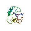
| |||||||||
|---|---|---|---|---|---|---|---|---|---|---|
| 1 |
| |||||||||
| NMR ensembles |
|
-Components
| #1: Protein | Mass: 10060.316 Da / Num. of mol.: 1 Source method: isolated from a genetically manipulated source Details: ATCC Source: (gene. exp.) Staphylococcus aureus subsp. aureus Mu50 (bacteria)Gene: purS, SAV1067 / Production host: References: UniProt: A0A0H3JUS1, phosphoribosylformylglycinamidine synthase |
|---|
-Experimental details
-Experiment
| Experiment | Method: SOLUTION NMR | ||||||||||||||||||||||||||||||||||||||||||||||||||||||||||||||||||
|---|---|---|---|---|---|---|---|---|---|---|---|---|---|---|---|---|---|---|---|---|---|---|---|---|---|---|---|---|---|---|---|---|---|---|---|---|---|---|---|---|---|---|---|---|---|---|---|---|---|---|---|---|---|---|---|---|---|---|---|---|---|---|---|---|---|---|---|
| NMR experiment |
|
 HSQC
HSQC- Sample preparation
| Details | Type: solution Contents: 1.2 mM 15N,13C Uniformly labeled Subunit PurS of phosphoribosylformylglycinamidine synthase, 20 mM sodium phosphate, 20 mM sodium chloride, 1 mM DTT, 0.03 % sodium azide, 0.15 mM DSS, 95% H2O/5% D2O Details: 1.2 mM protein in 20 mM sodium phosphate buffer pH 6.0, 20 mM sodium chloride, 1 mM DTT. Label: 15N,13C / Solvent system: 95% H2O/5% D2O | ||||||||||||||||||||||||||||
|---|---|---|---|---|---|---|---|---|---|---|---|---|---|---|---|---|---|---|---|---|---|---|---|---|---|---|---|---|---|
| Sample |
| ||||||||||||||||||||||||||||
| Sample conditions | Details: 1.2 mM protein in 20 mM sodium phosphate buffer pH 6.0, 20 mM sodium chloride, 1 mM DTT, NaN3 0.03%, DSS 0.15 mM Ionic strength: 20 mM / Label: 15N,13C Uniformly Labeled / pH: 6 / Pressure: 1 atm / Temperature: 298 K |
-NMR measurement
| NMR spectrometer | Type: Bruker AVANCE III HD / Manufacturer: Bruker / Model: AVANCE III HD / Field strength: 800 MHz |
|---|
- Processing
| NMR software |
| ||||||||||||||||||||||||||||||||||||||||
|---|---|---|---|---|---|---|---|---|---|---|---|---|---|---|---|---|---|---|---|---|---|---|---|---|---|---|---|---|---|---|---|---|---|---|---|---|---|---|---|---|---|
| Refinement | Method: na / Software ordinal: 6 | ||||||||||||||||||||||||||||||||||||||||
| NMR representative | Selection criteria: lowest energy | ||||||||||||||||||||||||||||||||||||||||
| NMR ensemble | Conformer selection criteria: structures with the lowest energy Conformers calculated total number: 100 / Conformers submitted total number: 18 |
