 Movie
Movie Controller

[English] 日本語
 Yorodumi
- PDB-8j2t: Glucosyl Transferase NbUGT72AY1 co-crystallized with UDP-2F gluco... -
+ Open data

- Basic information
| Entry | Database: PDB / ID: 8j2t | ||||||
|---|---|---|---|---|---|---|---|
| Title | Glucosyl Transferase NbUGT72AY1 co-crystallized with UDP-2F glucose and Scopoletin | ||||||
 Components Components | Glycosyltransferase | ||||||
 Keywords Keywords | TRANSFERASE / Glucosyl transferase | ||||||
| Function / homology | UDP-glycosyltransferase family, conserved site / UDP-glycosyltransferases signature. / UDP-glycosyltransferase activity / UDP-glucoronosyl and UDP-glucosyl transferase / UDP-glucuronosyl/UDP-glucosyltransferase / Transferases; Glycosyltransferases; Hexosyltransferases / 7-hydroxy-6-methoxy-2H-1-benzopyran-2-one / URIDINE-5'-DIPHOSPHATE-GLUCOSE / Glycosyltransferase Function and homology information Function and homology information | ||||||
| Biological species |  | ||||||
| Method |  X-RAY DIFFRACTION / SYNCHROTRON / MOLECULAR REPLACEMENT / Resolution: 3.15 Å X-RAY DIFFRACTION / SYNCHROTRON / MOLECULAR REPLACEMENT / Resolution: 3.15 Å | ||||||
 Authors Authors | Arold, S.T. / Hameed, U.F.S. | ||||||
| Funding support |  Saudi Arabia, 1items Saudi Arabia, 1items
| ||||||
 Citation Citation | Journal: To Be Published Title: Glucosyl Transferase NbUGT72AY1 co-crystallized with UDP-2F glucose and Scopoletin Authors: Arold, S.T. / Hameed, U.F.S. | ||||||
| History |
|
- Structure visualization
| Structure viewer | Molecule: MolmilJmol/JSmol |
|---|
- Downloads & links
-Download
| PDBx/mmCIF format | 8j2t.cif.gz | 199.8 KB | Display | PDBx/mmCIF format |
|---|---|---|---|---|
| PDB format | pdb8j2t.ent.gz | 158.3 KB | Display | PDB format |
| PDBx/mmJSON format | 8j2t.json.gz | Tree view | PDBx/mmJSON format | |
| Others |  Other downloads Other downloads |
-Validation report
| Arichive directory | https://data.pdbj.org/pub/pdb/validation_reports/j2/8j2tftp://data.pdbj.org/pub/pdb/validation_reports/j2/8j2t | HTTPS FTP |
|---|
-Related structure data
| Similar structure data |
|---|
-Links
 PDBj
PDBj
- Assembly
| Deposited unit | 
| ||||||||
|---|---|---|---|---|---|---|---|---|---|
| 1 | 
| ||||||||
| 2 | 
| ||||||||
| Unit cell |
|
-Components
| #1: Protein | Mass: 53945.941 Da / Num. of mol.: 2 Source method: isolated from a genetically manipulated source Source: (gene. exp.)  #2: Chemical | 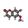  Mass: 192.168 Da / Num. of mol.: 2 / Source method: obtained synthetically / Formula: C10H8O4 / Feature type: SUBJECT OF INVESTIGATION Mass: 192.168 Da / Num. of mol.: 2 / Source method: obtained synthetically / Formula: C10H8O4 / Feature type: SUBJECT OF INVESTIGATION#3: Chemical |   Mass: 566.302 Da / Num. of mol.: 2 / Source method: obtained synthetically / Formula: C15H24N2O17P2 / Feature type: SUBJECT OF INVESTIGATION Mass: 566.302 Da / Num. of mol.: 2 / Source method: obtained synthetically / Formula: C15H24N2O17P2 / Feature type: SUBJECT OF INVESTIGATIONHas ligand of interest | Y | |
|---|
-Experimental details
-Experiment
| Experiment | Method: X-RAY DIFFRACTION / Number of used crystals: 1 |
|---|
- Sample preparation
| Crystal | Density Matthews: 2.75 Å3/Da / Density % sol: 55.22 % |
|---|---|
| Crystal grow | Temperature: 298 K / Method: vapor diffusion, hanging drop Details: 0.2 MLithium sulfate, 0.1 M Tris pH 8.5, 30% w/vPEG 4000 |
-Data collection
| Diffraction | Mean temperature: 100 K / Serial crystal experiment: N |
|---|---|
| Diffraction source | Source: SYNCHROTRON / Site: SOLEIL  / Beamline: PROXIMA 2 / Wavelength: 0.987 Å / Beamline: PROXIMA 2 / Wavelength: 0.987 Å |
| Detector | Type: DECTRIS EIGER X 9M / Detector: PIXEL / Date: Dec 10, 2022 |
| Radiation | Protocol: SINGLE WAVELENGTH / Monochromatic (M) / Laue (L): M / Scattering type: x-ray |
| Radiation wavelength | Wavelength: 0.987 Å / Relative weight: 1 |
| Reflection | Resolution: 3.08→46.9 Å / Num. obs: 18992 / % possible obs: 97.2 % / Redundancy: 3.6 % / CC1/2: 0.984 / Net I/σ(I): 4.9 |
| Reflection shell | Resolution: 3.08→3.16 Å / Num. unique obs: 3327 / CC1/2: 0.464 |
- Processing
| Software |
| ||||||||||||||||||||
|---|---|---|---|---|---|---|---|---|---|---|---|---|---|---|---|---|---|---|---|---|---|
| Refinement | Method to determine structure: MOLECULAR REPLACEMENT / Resolution: 3.15→46.83 Å / SU B: 27.782 / SU ML: 0.454 / Cross valid method: THROUGHOUT / ESU R Free: 0.531 / Details: HYDROGENS HAVE BEEN ADDED IN THE RIDING POSITIONS
| ||||||||||||||||||||
| Displacement parameters | Biso mean: 77.21 Å2
| ||||||||||||||||||||
| Refinement step | Cycle: LAST / Resolution: 3.15→46.83 Å
| ||||||||||||||||||||
| LS refinement shell | Resolution: 3.152→3.234 Å
|
