 Movie
Movie Controller


+ Open data

- Basic information
| Entry | Database: PDB / ID: 8j2f | ||||||
|---|---|---|---|---|---|---|---|
| Title | Human neutral shpingomyelinase | ||||||
 Components Components | Sphingomyelin phosphodiesterase 2 | ||||||
 Keywords Keywords | MEMBRANE PROTEIN / enzyme | ||||||
| Function / homology |  Function and homology information Function and homology informationsphingomyelin metabolic process / sphingomyelin catabolic process / sphingomyelin phosphodiesterase / Ceramide signalling / sphingomyelin phosphodiesterase activity / Glycosphingolipid catabolism / phosphoric diester hydrolase activity / sphingolipid catabolic process / ceramide biosynthetic process / TNFR1-mediated ceramide production ...sphingomyelin metabolic process / sphingomyelin catabolic process / sphingomyelin phosphodiesterase / Ceramide signalling / sphingomyelin phosphodiesterase activity / Glycosphingolipid catabolism / phosphoric diester hydrolase activity / sphingolipid catabolic process / ceramide biosynthetic process / TNFR1-mediated ceramide production / response to mechanical stimulus / cell periphery / caveola / intracellular signal transduction / endoplasmic reticulum / metal ion binding / plasma membrane Similarity search - Function | ||||||
| Biological species |  Homo sapiens (human) Homo sapiens (human) | ||||||
| Method | ELECTRON MICROSCOPY / single particle reconstruction / cryo EM / Resolution: 3.07 Å | ||||||
 Authors Authors | Zhang, S.S. | ||||||
| Funding support |  China, 1items China, 1items
| ||||||
 Citation Citation | Journal: Nat Commun / Year: 2023 Title: Molecular basis for the catalytic mechanism of human neutral sphingomyelinases 1 (hSMPD2). Authors: Jingbo Yi / Boya Qi / Jian Yin / Ruochong Li / Xudong Chen / Junhan Hu / Guohui Li / Sensen Zhang / Yuebin Zhang / Maojun Yang / Abstract: Enzymatic breakdown of sphingomyelin by sphingomyelinase (SMase) is the main source of the membrane lipids, ceramides, which are involved in many cellular physiological processes. However, the full- ...Enzymatic breakdown of sphingomyelin by sphingomyelinase (SMase) is the main source of the membrane lipids, ceramides, which are involved in many cellular physiological processes. However, the full-length structure of human neutral SMase has not been resolved; therefore, its catalytic mechanism remains unknown. Here, we resolve the structure of human full-length neutral SMase, sphingomyelinase 1 (SMPD2), which reveals that C-terminal transmembrane helices contribute to dimeric architecture of hSMPD2 and that D111 - K116 loop domain is essential for substrate hydrolysis. Coupled with molecular docking, we clarify the binding pose of sphingomyelin, and site-directed mutagenesis further confirms key residues responsible for sphingomyelin binding. Hybrid quantum mechanics/molecular mechanics (QM/MM) molecular dynamic (MD) simulations are utilized to elaborate the catalysis of hSMPD2 with the reported in vitro substrates, sphingomyelin and lyso-platelet activating fator (lyso-PAF). Our study provides mechanistic details that enhance our knowledge of lipid metabolism and may lead to an improved understanding of ceramide in disease and in cancer treatment. | ||||||
| History |
|
- Structure visualization
| Structure viewer | Molecule: MolmilJmol/JSmol |
|---|
- Downloads & links
-Download
| PDBx/mmCIF format | 8j2f.cif.gz | 144.3 KB | Display | PDBx/mmCIF format |
|---|---|---|---|---|
| PDB format | pdb8j2f.ent.gz | 113.1 KB | Display | PDB format |
| PDBx/mmJSON format | 8j2f.json.gz | Tree view | PDBx/mmJSON format | |
| Others |  Other downloads Other downloads |
-Validation report
| Arichive directory | https://data.pdbj.org/pub/pdb/validation_reports/j2/8j2fftp://data.pdbj.org/pub/pdb/validation_reports/j2/8j2f | HTTPS FTP |
|---|
-Related structure data
| Related structure data |  35948MC M: map data used to model this data C: citing same article ( |
|---|---|
| Similar structure data |
-Links
 PDBj
PDBj


- Assembly
| Deposited unit | 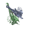
|
|---|---|
| 1 |
|
-Components
| #1: Protein | Mass: 47702.508 Da / Num. of mol.: 2 Source method: isolated from a genetically manipulated source Source: (gene. exp.) Homo sapiens (human) / Gene: SMPD2 / Production host: Homo sapiens (human)References: UniProt: O60906, sphingomyelin phosphodiesterase #2: Chemical |   Mass: 24.305 Da / Num. of mol.: 2 / Source method: obtained synthetically / Formula: Mg Mass: 24.305 Da / Num. of mol.: 2 / Source method: obtained synthetically / Formula: Mg#3: Chemical | 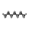  Mass: 100.202 Da / Num. of mol.: 2 / Source method: obtained synthetically / Formula: C7H16 Mass: 100.202 Da / Num. of mol.: 2 / Source method: obtained synthetically / Formula: C7H16#4: Chemical |   Mass: 198.388 Da / Num. of mol.: 2 / Source method: obtained synthetically / Formula: C14H30 Mass: 198.388 Da / Num. of mol.: 2 / Source method: obtained synthetically / Formula: C14H30Has ligand of interest | N | |
|---|
-Experimental details
-Experiment
| Experiment | Method: ELECTRON MICROSCOPY |
|---|---|
| EM experiment | Aggregation state: PARTICLE / 3D reconstruction method: single particle reconstruction |
- Sample preparation
| Component | Name: human neutral sphingomyelinases / Type: COMPLEX / Entity ID: #1 / Source: MULTIPLE SOURCES |
|---|---|
| Source (natural) | Organism: Homo sapiens (human) |
| Source (recombinant) | Organism: Homo sapiens (human) |
| Buffer solution | pH: 7.2 |
| Specimen | Embedding applied: NO / Shadowing applied: NO / Staining applied: NO / Vitrification applied: YES |
| Vitrification | Cryogen name: ETHANE |
- Electron microscopy imaging
| Experimental equipment |  Model: Titan Krios / Image courtesy: FEI Company |
|---|---|
| Microscopy | Model: FEI TITAN KRIOS |
| Electron gun | Electron source:  FIELD EMISSION GUN / Accelerating voltage: 300 kV / Illumination mode: FLOOD BEAM FIELD EMISSION GUN / Accelerating voltage: 300 kV / Illumination mode: FLOOD BEAM |
| Electron lens | Mode: BRIGHT FIELD / Nominal defocus max: 2000 nm / Nominal defocus min: 1200 nm |
| Image recording | Electron dose: 50 e/Å2 / Film or detector model: GATAN K3 BIOQUANTUM (6k x 4k) |
- Processing
| CTF correction | Type: PHASE FLIPPING AND AMPLITUDE CORRECTION |
|---|---|
| 3D reconstruction | Resolution: 3.07 Å / Resolution method: FSC 0.143 CUT-OFF / Num. of particles: 220000 / Symmetry type: POINT |
