 Movie
Movie Controller


+ Open data

- Basic information
| Entry | Database: PDB / ID: 8j1g | ||||||
|---|---|---|---|---|---|---|---|
| Title | Structure of amino acid dehydrogenase in complex with NADPH | ||||||
 Components Components | Ornithine cyclodeaminase family protein | ||||||
 Keywords Keywords | OXIDOREDUCTASE / dehydrogenase | ||||||
| Function / homology | Ornithine cyclodeaminase/mu-crystallin / Ornithine cyclodeaminase, N-terminal / Ornithine cyclodeaminase/mu-crystallin family / NAD(P)-binding domain superfamily / ARGININE / Chem-NDP / Ornithine cyclodeaminase family protein Function and homology information Function and homology information | ||||||
| Biological species |  Pseudomonas veronii (bacteria) Pseudomonas veronii (bacteria) | ||||||
| Method |  X-RAY DIFFRACTION / SYNCHROTRON / MOLECULAR REPLACEMENT / Resolution: 2.5 Å X-RAY DIFFRACTION / SYNCHROTRON / MOLECULAR REPLACEMENT / Resolution: 2.5 Å | ||||||
 Authors Authors | Sakuraba, H. / Ohshima, T. | ||||||
| Funding support |  Japan, 1items Japan, 1items
| ||||||
 Citation Citation | Journal: Int.J.Biol.Macromol. / Year: 2023 Title: First crystal structure of an NADP + -dependent l-arginine dehydrogenase belonging to the mu-crystallin family. Authors: Kawakami, R. / Takami, N. / Hayashi, J. / Yoneda, K. / Ohmori, T. / Ohshima, T. / Sakuraba, H. | ||||||
| History |
|
- Structure visualization
| Structure viewer | Molecule: MolmilJmol/JSmol |
|---|
- Downloads & links
-Download
| PDBx/mmCIF format | 8j1g.cif.gz | 483.7 KB | Display | PDBx/mmCIF format |
|---|---|---|---|---|
| PDB format | pdb8j1g.ent.gz | 400.8 KB | Display | PDB format |
| PDBx/mmJSON format | 8j1g.json.gz | Tree view | PDBx/mmJSON format | |
| Others |  Other downloads Other downloads |
-Validation report
| Summary document | 8j1g_validation.pdf.gz | 2.4 MB | Display | wwPDB validaton report |
|---|---|---|---|---|
| Full document | 8j1g_full_validation.pdf.gz | 2.4 MB | Display | |
| Data in XML | 8j1g_validation.xml.gz | 46.6 KB | Display | |
| Data in CIF | 8j1g_validation.cif.gz | 61.7 KB | Display | |
| Arichive directory | https://data.pdbj.org/pub/pdb/validation_reports/j1/8j1gftp://data.pdbj.org/pub/pdb/validation_reports/j1/8j1g | HTTPS FTP |
-Related structure data

-Links
 PDBj
PDBj


- Assembly
| Deposited unit | 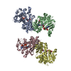
| ||||||||
|---|---|---|---|---|---|---|---|---|---|
| 1 | 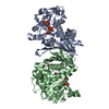
| ||||||||
| 2 | 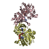
| ||||||||
| Unit cell |
|
-Components
| #1: Protein | Mass: 36036.043 Da / Num. of mol.: 4 Source method: isolated from a genetically manipulated source Source: (gene. exp.) Pseudomonas veronii (bacteria) / Gene: HBO43_18005 / Production host: #2: Chemical | ChemComp-NDP /   Mass: 745.421 Da / Num. of mol.: 4 / Source method: obtained synthetically / Formula: C21H30N7O17P3 / Feature type: SUBJECT OF INVESTIGATION Mass: 745.421 Da / Num. of mol.: 4 / Source method: obtained synthetically / Formula: C21H30N7O17P3 / Feature type: SUBJECT OF INVESTIGATION#3: Chemical | ChemComp-ARG /   Type: L-peptide linking / Mass: 175.209 Da / Num. of mol.: 4 / Source method: obtained synthetically / Formula: C6H15N4O2 Type: L-peptide linking / Mass: 175.209 Da / Num. of mol.: 4 / Source method: obtained synthetically / Formula: C6H15N4O2#4: Chemical |   Mass: 62.068 Da / Num. of mol.: 2 / Source method: obtained synthetically / Formula: C2H6O2 / Feature type: SUBJECT OF INVESTIGATION Mass: 62.068 Da / Num. of mol.: 2 / Source method: obtained synthetically / Formula: C2H6O2 / Feature type: SUBJECT OF INVESTIGATION#5: Water | ChemComp-HOH / |  Mass: 18.015 Da / Num. of mol.: 45 / Source method: isolated from a natural source / Formula: H2O Mass: 18.015 Da / Num. of mol.: 45 / Source method: isolated from a natural source / Formula: H2OHas ligand of interest | Y | |
|---|
-Experimental details
-Experiment
| Experiment | Method: X-RAY DIFFRACTION / Number of used crystals: 1 |
|---|
- Sample preparation
| Crystal | Density Matthews: 2.95 Å3/Da / Density % sol: 58.33 % |
|---|---|
| Crystal grow | Temperature: 293 K / Method: vapor diffusion, sitting drop / pH: 4.6 Details: PEG 4000, Na acetate buffer, glycerol, NADPH, arginine |
-Data collection
| Diffraction | Mean temperature: 100 K / Serial crystal experiment: N |
|---|---|
| Diffraction source | Source: SYNCHROTRON / Site: Photon Factory / Beamline: BL-5A / Wavelength: 1 Å |
| Detector | Type: DECTRIS PILATUS 6M / Detector: PIXEL / Date: Dec 16, 2022 |
| Radiation | Monochromator: Si / Protocol: SINGLE WAVELENGTH / Monochromatic (M) / Laue (L): M / Scattering type: x-ray |
| Radiation wavelength | Wavelength: 1 Å / Relative weight: 1 |
| Reflection | Resolution: 2.5→47.8 Å / Num. obs: 56308 / % possible obs: 98.7 % / Redundancy: 7 % / CC1/2: 0.999 / Rmerge(I) obs: 0.063 / Rpim(I) all: 0.026 / Rrim(I) all: 0.068 / Χ2: 0.99 / Net I/σ(I): 18.9 |
| Reflection shell | Resolution: 2.5→2.57 Å / % possible obs: 98.1 % / Redundancy: 6.7 % / Rmerge(I) obs: 1.2 / Num. measured all: 31137 / Num. unique obs: 4614 / CC1/2: 0.657 / Rpim(I) all: 0.495 / Rrim(I) all: 1.3 / Χ2: 0.96 / Net I/σ(I) obs: 1.8 |
- Processing
| Software |
| ||||||||||||||||||||||||||||||||||||||||||||||||||||||||||||||||||||||||||||||||||||||||||||||||||||||||||||||||||||||||||||||||||||||||||||||||||||||||||||||||||||||||||||||||||||||
|---|---|---|---|---|---|---|---|---|---|---|---|---|---|---|---|---|---|---|---|---|---|---|---|---|---|---|---|---|---|---|---|---|---|---|---|---|---|---|---|---|---|---|---|---|---|---|---|---|---|---|---|---|---|---|---|---|---|---|---|---|---|---|---|---|---|---|---|---|---|---|---|---|---|---|---|---|---|---|---|---|---|---|---|---|---|---|---|---|---|---|---|---|---|---|---|---|---|---|---|---|---|---|---|---|---|---|---|---|---|---|---|---|---|---|---|---|---|---|---|---|---|---|---|---|---|---|---|---|---|---|---|---|---|---|---|---|---|---|---|---|---|---|---|---|---|---|---|---|---|---|---|---|---|---|---|---|---|---|---|---|---|---|---|---|---|---|---|---|---|---|---|---|---|---|---|---|---|---|---|---|---|---|---|
| Refinement | Method to determine structure: MOLECULAR REPLACEMENT / Resolution: 2.5→47.8 Å / Cor.coef. Fo:Fc: 0.95 / Cor.coef. Fo:Fc free: 0.923 / SU B: 38.301 / SU ML: 0.349 / Cross valid method: THROUGHOUT / ESU R: 0.452 / ESU R Free: 0.295 / Stereochemistry target values: MAXIMUM LIKELIHOOD / Details: HYDROGENS HAVE BEEN ADDED IN THE RIDING POSITIONS
| ||||||||||||||||||||||||||||||||||||||||||||||||||||||||||||||||||||||||||||||||||||||||||||||||||||||||||||||||||||||||||||||||||||||||||||||||||||||||||||||||||||||||||||||||||||||
| Solvent computation | Ion probe radii: 0.8 Å / Shrinkage radii: 0.8 Å / VDW probe radii: 1.2 Å / Solvent model: MASK | ||||||||||||||||||||||||||||||||||||||||||||||||||||||||||||||||||||||||||||||||||||||||||||||||||||||||||||||||||||||||||||||||||||||||||||||||||||||||||||||||||||||||||||||||||||||
| Displacement parameters | Biso mean: 80.876 Å2
| ||||||||||||||||||||||||||||||||||||||||||||||||||||||||||||||||||||||||||||||||||||||||||||||||||||||||||||||||||||||||||||||||||||||||||||||||||||||||||||||||||||||||||||||||||||||
| Refinement step | Cycle: 1 / Resolution: 2.5→47.8 Å
| ||||||||||||||||||||||||||||||||||||||||||||||||||||||||||||||||||||||||||||||||||||||||||||||||||||||||||||||||||||||||||||||||||||||||||||||||||||||||||||||||||||||||||||||||||||||
| Refine LS restraints |
|