 Movie
Movie Controller


+ Open data

- Basic information
| Entry | Database: PDB / ID: 8iyf | ||||||
|---|---|---|---|---|---|---|---|
| Title | Structure of VldE-H182A in complex with GDP | ||||||
 Components Components | Validamine 7-phosphate valienyltransferase | ||||||
 Keywords Keywords | TRANSFERASE / Pseudoglycosyltransferase / validamine | ||||||
| Function / homology | validamine 7-phosphate valienyltransferase / alpha,alpha-trehalose-phosphate synthase (UDP-forming) activity / Glycosyl transferase, family 20 / Glycosyltransferase family 20 / trehalose biosynthetic process / antibiotic biosynthetic process / GUANOSINE-5'-DIPHOSPHATE / Validamine 7-phosphate valienyltransferase Function and homology information Function and homology information | ||||||
| Biological species |  Streptomyces hygroscopicus subsp. limoneus (bacteria) Streptomyces hygroscopicus subsp. limoneus (bacteria) | ||||||
| Method |  X-RAY DIFFRACTION / SYNCHROTRON / MOLECULAR REPLACEMENT / Resolution: 1.9 Å X-RAY DIFFRACTION / SYNCHROTRON / MOLECULAR REPLACEMENT / Resolution: 1.9 Å | ||||||
 Authors Authors | Wong, C.P. / Mori, T. / Mahmud, T. / Abe, I. | ||||||
| Funding support | 1items
| ||||||
 Citation Citation | Journal: To Be Published Title: Structure of VldE-H182A in complex with GDP Authors: Wong, C.P. / Mori, T. / Mahmud, T. / Abe, I. | ||||||
| History |
|
- Structure visualization
| Structure viewer | Molecule: MolmilJmol/JSmol |
|---|
- Downloads & links
-Download
| PDBx/mmCIF format | 8iyf.cif.gz | 209.9 KB | Display | PDBx/mmCIF format |
|---|---|---|---|---|
| PDB format | pdb8iyf.ent.gz | 162.2 KB | Display | PDB format |
| PDBx/mmJSON format | 8iyf.json.gz | Tree view | PDBx/mmJSON format | |
| Others |  Other downloads Other downloads |
-Validation report
| Arichive directory | https://data.pdbj.org/pub/pdb/validation_reports/iy/8iyfftp://data.pdbj.org/pub/pdb/validation_reports/iy/8iyf | HTTPS FTP |
|---|
-Related structure data
| Similar structure data |
|---|
-Links
 PDBj
PDBj- Assembly
| Deposited unit | 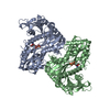
| ||||||||||||
|---|---|---|---|---|---|---|---|---|---|---|---|---|---|
| 1 |
| ||||||||||||
| Unit cell |
|
-Components
| #1: Protein | Mass: 54939.465 Da / Num. of mol.: 2 / Mutation: H182A Source method: isolated from a genetically manipulated source Source: (gene. exp.) Streptomyces hygroscopicus subsp. limoneus (bacteria)Gene: vldE, SHL15_8006 / Production host: References: UniProt: Q15JG1, validamine 7-phosphate valienyltransferase #2: Chemical |   Type: RNA linking / Mass: 443.201 Da / Num. of mol.: 2 / Source method: obtained synthetically / Formula: C10H15N5O11P2 / Feature type: SUBJECT OF INVESTIGATION / Comment: GDP, energy-carrying molecule*YM Type: RNA linking / Mass: 443.201 Da / Num. of mol.: 2 / Source method: obtained synthetically / Formula: C10H15N5O11P2 / Feature type: SUBJECT OF INVESTIGATION / Comment: GDP, energy-carrying molecule*YM#3: Water | ChemComp-HOH / |  Mass: 18.015 Da / Num. of mol.: 588 / Source method: isolated from a natural source / Formula: H2O Mass: 18.015 Da / Num. of mol.: 588 / Source method: isolated from a natural source / Formula: H2OHas ligand of interest | Y | |
|---|
-Experimental details
-Experiment
| Experiment | Method: X-RAY DIFFRACTION / Number of used crystals: 1 |
|---|
- Sample preparation
| Crystal | Density Matthews: 2.24 Å3/Da / Density % sol: 45.08 % |
|---|---|
| Crystal grow | Temperature: 293 K / Method: vapor diffusion, sitting drop / Details: 100 mM Tris-HCl, pH8.5, 26% PEG 4000 |
-Data collection
| Diffraction | Mean temperature: 100 K / Serial crystal experiment: N |
|---|---|
| Diffraction source | Source: SYNCHROTRON / Site: NSRRC  / Beamline: TPS 05A / Wavelength: 1 Å / Beamline: TPS 05A / Wavelength: 1 Å |
| Detector | Type: RAYONIX MX300-HS / Detector: CCD / Date: Aug 23, 2017 |
| Radiation | Protocol: SINGLE WAVELENGTH / Monochromatic (M) / Laue (L): M / Scattering type: x-ray |
| Radiation wavelength | Wavelength: 1 Å / Relative weight: 1 |
| Reflection | Resolution: 1.9→48.01 Å / Num. obs: 74391 / % possible obs: 97.9 % / Redundancy: 4.4 % / Biso Wilson estimate: 26.21 Å2 / CC1/2: 0.992 / Rmerge(I) obs: 0.09 / Net I/σ(I): 10.3 |
| Reflection shell | Resolution: 1.9→1.94 Å / Rmerge(I) obs: 0.518 / Mean I/σ(I) obs: 2.3 / Num. unique obs: 4500 / CC1/2: 0.805 |
- Processing
| Software |
| |||||||||||||||||||||||||||||||||||||||||||||||||||||||||||||||||||||||||||||||||||||||||||||||||||||||||
|---|---|---|---|---|---|---|---|---|---|---|---|---|---|---|---|---|---|---|---|---|---|---|---|---|---|---|---|---|---|---|---|---|---|---|---|---|---|---|---|---|---|---|---|---|---|---|---|---|---|---|---|---|---|---|---|---|---|---|---|---|---|---|---|---|---|---|---|---|---|---|---|---|---|---|---|---|---|---|---|---|---|---|---|---|---|---|---|---|---|---|---|---|---|---|---|---|---|---|---|---|---|---|---|---|---|---|
| Refinement | Method to determine structure: MOLECULAR REPLACEMENT / Resolution: 1.9→48.01 Å / SU ML: 0.2376 / Cross valid method: FREE R-VALUE / σ(F): 1.39 / Phase error: 25.4978 Stereochemistry target values: GeoStd + Monomer Library + CDL v1.2
| |||||||||||||||||||||||||||||||||||||||||||||||||||||||||||||||||||||||||||||||||||||||||||||||||||||||||
| Solvent computation | Shrinkage radii: 0.9 Å / VDW probe radii: 1.11 Å / Solvent model: FLAT BULK SOLVENT MODEL | |||||||||||||||||||||||||||||||||||||||||||||||||||||||||||||||||||||||||||||||||||||||||||||||||||||||||
| Displacement parameters | Biso mean: 29.83 Å2 | |||||||||||||||||||||||||||||||||||||||||||||||||||||||||||||||||||||||||||||||||||||||||||||||||||||||||
| Refinement step | Cycle: LAST / Resolution: 1.9→48.01 Å
| |||||||||||||||||||||||||||||||||||||||||||||||||||||||||||||||||||||||||||||||||||||||||||||||||||||||||
| Refine LS restraints |
| |||||||||||||||||||||||||||||||||||||||||||||||||||||||||||||||||||||||||||||||||||||||||||||||||||||||||
| LS refinement shell |
|
