 Movie
Movie Controller


+ Open data

- Basic information
| Entry | Database: PDB / ID: 8ivn | ||||||
|---|---|---|---|---|---|---|---|
| Title | crystal structure of SulE mutant | ||||||
 Components Components | Alpha/beta fold hydrolase | ||||||
 Keywords Keywords | HYDROLASE / Complex / SulE / mutant | ||||||
| Function / homology |  Function and homology information Function and homology informationHydrolases; Acting on ester bonds; Carboxylic-ester hydrolases / hydrolase activity Similarity search - Function | ||||||
| Biological species |  Hansschlegelia zhihuaiae (bacteria) Hansschlegelia zhihuaiae (bacteria) | ||||||
| Method |  X-RAY DIFFRACTION / SYNCHROTRON / MOLECULAR REPLACEMENT / Resolution: 1.5 Å X-RAY DIFFRACTION / SYNCHROTRON / MOLECULAR REPLACEMENT / Resolution: 1.5 Å | ||||||
 Authors Authors | Liu, B. / He, J. / Ran, T. / Wang, W. | ||||||
| Funding support |  China, 1items China, 1items
| ||||||
 Citation Citation | Journal: Nat Commun / Year: 2023 Title: Crystal structures of herbicide-detoxifying esterase reveal a lid loop affecting substrate binding and activity. Authors: Liu, B. / Wang, W. / Qiu, J. / Huang, X. / Qiu, S. / Bao, Y. / Xu, S. / Ruan, L. / Ran, T. / He, J. | ||||||
| History |
|
- Structure visualization
| Structure viewer | Molecule: MolmilJmol/JSmol |
|---|
- Downloads & links
-Download
| PDBx/mmCIF format | 8ivn.cif.gz | 173.5 KB | Display | PDBx/mmCIF format |
|---|---|---|---|---|
| PDB format | pdb8ivn.ent.gz | 132.4 KB | Display | PDB format |
| PDBx/mmJSON format | 8ivn.json.gz | Tree view | PDBx/mmJSON format | |
| Others |  Other downloads Other downloads |
-Validation report
| Arichive directory | https://data.pdbj.org/pub/pdb/validation_reports/iv/8ivnftp://data.pdbj.org/pub/pdb/validation_reports/iv/8ivn | HTTPS FTP |
|---|
-Related structure data
| Related structure data | 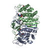 7y0lC  7yd2C  8golSC  8goyC 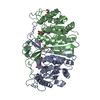 8gp0C 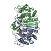 8iveC 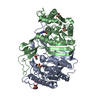 8ivmC  8ivsC 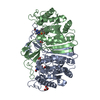 8ivtC 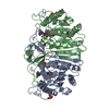 8iw3C 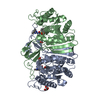 8iw6C 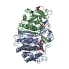 8j7gC  8j7jC  8j7kC S: Starting model for refinement C: citing same article ( |
|---|---|
| Similar structure data |
-Links
 PDBj
PDBj


- Assembly
| Deposited unit | 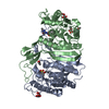
| ||||||||||||
|---|---|---|---|---|---|---|---|---|---|---|---|---|---|
| 1 |
| ||||||||||||
| Unit cell |
|
-Components
| #1: Protein | Mass: 41092.234 Da / Num. of mol.: 2 / Mutation: S209A,H333A Source method: isolated from a genetically manipulated source Source: (gene. exp.) Hansschlegelia zhihuaiae (bacteria) / Gene: EK403_17710 / Production host: #2: Chemical | 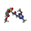  Mass: 381.364 Da / Num. of mol.: 3 / Source method: obtained synthetically / Formula: C14H15N5O6S / Feature type: SUBJECT OF INVESTIGATION Mass: 381.364 Da / Num. of mol.: 3 / Source method: obtained synthetically / Formula: C14H15N5O6S / Feature type: SUBJECT OF INVESTIGATION#3: Chemical |   Mass: 150.087 Da / Num. of mol.: 2 / Source method: obtained synthetically / Formula: C4H6O6 / Feature type: SUBJECT OF INVESTIGATION Mass: 150.087 Da / Num. of mol.: 2 / Source method: obtained synthetically / Formula: C4H6O6 / Feature type: SUBJECT OF INVESTIGATION#4: Chemical |   Mass: 92.094 Da / Num. of mol.: 2 / Source method: obtained synthetically / Formula: C3H8O3 Mass: 92.094 Da / Num. of mol.: 2 / Source method: obtained synthetically / Formula: C3H8O3#5: Water | ChemComp-HOH / |  Mass: 18.015 Da / Num. of mol.: 851 / Source method: isolated from a natural source / Formula: H2O Mass: 18.015 Da / Num. of mol.: 851 / Source method: isolated from a natural source / Formula: H2OHas ligand of interest | Y | |
|---|
-Experimental details
-Experiment
| Experiment | Method: X-RAY DIFFRACTION / Number of used crystals: 1 |
|---|
- Sample preparation
| Crystal | Density Matthews: 2.48 Å3/Da / Density % sol: 50.45 % |
|---|---|
| Crystal grow | Temperature: 277 K / Method: vapor diffusion, sitting drop / Details: PEG |
-Data collection
| Diffraction | Mean temperature: 100 K / Serial crystal experiment: N |
|---|---|
| Diffraction source | Source: SYNCHROTRON / Site: SSRF / Beamline: BL18U1 / Wavelength: 0.9791 Å |
| Detector | Type: DECTRIS PILATUS 6M / Detector: PIXEL / Date: Jan 1, 2023 |
| Radiation | Protocol: SINGLE WAVELENGTH / Monochromatic (M) / Laue (L): M / Scattering type: x-ray |
| Radiation wavelength | Wavelength: 0.9791 Å / Relative weight: 1 |
| Reflection | Resolution: 1.5→42.03 Å / Num. obs: 126161 / % possible obs: 98.9 % / Redundancy: 6 % / CC1/2: 0.992 / Rpim(I) all: 0.04 / Net I/σ(I): 16.4 |
| Reflection shell | Resolution: 1.5→1.54 Å / Num. unique obs: 8460 / CC1/2: 0.698 / Rpim(I) all: 0.395 |
- Processing
| Software |
| ||||||||||||||||||||||||
|---|---|---|---|---|---|---|---|---|---|---|---|---|---|---|---|---|---|---|---|---|---|---|---|---|---|
| Refinement | Method to determine structure: MOLECULAR REPLACEMENT Starting model: 8GOL Resolution: 1.5→40.78 Å / Cross valid method: FREE R-VALUE Stereochemistry target values: GeoStd + Monomer Library + CDL v1.2
| ||||||||||||||||||||||||
| Displacement parameters | Biso mean: 17.94 Å2 | ||||||||||||||||||||||||
| Refinement step | Cycle: LAST / Resolution: 1.5→40.78 Å
| ||||||||||||||||||||||||
| Refine LS restraints |
|
