[English] 日本語
Yorodumi
- PDB-8iqc: Crystal structure of AsfvPrimPol N-terminal Prim/Pol domain in co... -

+
Open data


ID or keywords:

Loading...

-
Basic information

Entry
Database: PDB / ID: 8iqc
TitleCrystal structure of AsfvPrimPol N-terminal Prim/Pol domain in complex with Mn2+
ComponentsPutative primase C962R
KeywordsDNA BINDING PROTEIN / polymerase / primase / PrimPol
Function / homology
Function and homology information


hydrolase activity, acting on acid anhydrides / helicase activity / DNA replication / ATP binding / metal ion binding
Similarity search - Function
Primase, C-terminal 2 / Primase C terminal 2 (PriCT-2) / Domain of unknown function DUF5906 / Family of unknown function (DUF5906) / Bacteriophage/plasmid primase, P4, C-terminal / D5 N terminal like / Helicase, superfamily 3, DNA virus / Superfamily 3 helicase of DNA viruses domain profile. / P-loop containing nucleoside triphosphate hydrolase
Similarity search - Domain/homology
: / Putative primase C962R
Similarity search - Component
Biological speciesAfrican swine fever virus BA71V
MethodX-RAY DIFFRACTION / SYNCHROTRON / MOLECULAR REPLACEMENT / Resolution: 2 Å
AuthorsShao, Z.W. / Gan, J.H.
Funding support China, 1items
OrganizationGrant numberCountry
National Natural Science Foundation of China (NSFC) China
Citation
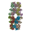
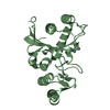
Journal: Nucleic Acids Res / Year: 2023
Title: Structures and implications of the C962R protein of African swine fever virus.
Authors: Zhiwei Shao / Shichen Su / Jie Yang / Weizhen Zhang / Yanqing Gao / Xin Zhao / Yixi Zhang / Qiyuan Shao / Chulei Cao / Huili Li / Hehua Liu / Jinru Zhang / Jinzhong Lin / Jinbiao Ma / Jianhua Gan /
Abstract: African swine fever virus (ASFV) is highly contagious and can cause lethal disease in pigs. Although it has been extensively studied in the past, no vaccine or other useful treatment against ASFV is ...African swine fever virus (ASFV) is highly contagious and can cause lethal disease in pigs. Although it has been extensively studied in the past, no vaccine or other useful treatment against ASFV is available. The genome of ASFV encodes more than 170 proteins, but the structures and functions for the majority of the proteins remain elusive, which hindered our understanding on the life cycle of ASFV and the development of ASFV-specific inhibitors. Here, we report the structural and biochemical studies of the highly conserved C962R protein of ASFV, showing that C962R is a multidomain protein. The N-terminal AEP domain is responsible for the DNA polymerization activity, whereas the DNA unwinding activity is catalyzed by the central SF3 helicase domain. The middle PriCT2 and D5_N domains and the C-terminal Tail domain all contribute to the DNA unwinding activity of C962R. C962R preferentially works on forked DNA, and likely functions in Base-excision repair (BER) or other repair pathway in ASFV. Although it is not essential for the replication of ASFV, C962R can serve as a model and provide mechanistic insight into the replicative primase proteins from many other species, such as nitratiruptor phage NrS-1, vaccinia virus (VACV) and other viruses.
History
DepositionMar 16, 2023Deposition site: PDBJ / Processing site: PDBJ
Revision 1.0Jul 26, 2023Provider: repository / Type: Initial release
Revision 1.1Nov 29, 2023Group: Data collection / Database references
Category: chem_comp_atom / chem_comp_bond ...chem_comp_atom / chem_comp_bond / citation / citation_author
Item: _citation.country / _citation.journal_abbrev ..._citation.country / _citation.journal_abbrev / _citation.journal_id_ASTM / _citation.journal_id_CSD / _citation.journal_id_ISSN / _citation.journal_volume / _citation.page_first / _citation.page_last / _citation.pdbx_database_id_DOI / _citation.pdbx_database_id_PubMed / _citation.title / _citation.year

-
Structure visualization

Structure viewerMolecule:
MolmilJmol/JSmol

Downloads & links

-
Assembly

Deposited unit
A: Putative primase C962R
B: Putative primase C962R
hetero molecules


Theoretical massNumber of molelcules
Total (without water)56,9576
Polymers56,7372
Non-polymers2204
Water2,936163
1
A: Putative primase C962R
hetero molecules


Theoretical massNumber of molelcules
Total (without water)28,4783
Polymers28,3691
Non-polymers1102
Water181
TypeNameSymmetry operationNumber
identity operation1_555x,y,z1
2
B: Putative primase C962R
hetero molecules


Theoretical massNumber of molelcules
Total (without water)28,4783
Polymers28,3691
Non-polymers1102
Water181
TypeNameSymmetry operationNumber
identity operation1_555x,y,z1
Unit cell
Length a, b, c (Å)128.424, 43.922, 87.584
Angle α, β, γ (deg.)90.00, 93.29, 90.00
Int Tables number5
Space group name H-MC121

-
Components

#1: Protein Putative primase C962R


Mass: 28368.506 Da / Num. of mol.: 2 / Fragment: N-terminal Prim/Pol domain
Source method: isolated from a genetically manipulated source
Source: (gene. exp.) African swine fever virus BA71V / Gene: BA71V-C962R, C962R / Production host: Escherichia coli (E. coli) / References: UniProt: A0A0C5B022
#2: Chemical
ChemComp-MN / MANGANESE (II) ION


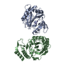
Mass: 54.938 Da / Num. of mol.: 4 / Source method: obtained synthetically / Formula: Mn / Feature type: SUBJECT OF INVESTIGATION
#3: Water ChemComp-HOH / water


Mass: 18.015 Da / Num. of mol.: 163 / Source method: isolated from a natural source / Formula: H2O
Has ligand of interestY

-
Experimental details

-
Experiment

ExperimentMethod: X-RAY DIFFRACTION / Number of used crystals: 1

-
Sample preparation

CrystalDensity Matthews: 2.17 Å3/Da / Density % sol: 43.4 %
Crystal growTemperature: 291 K / Method: vapor diffusion, sitting drop
Details: 0.1 M Bis-Tris propane pH 7.5, 25% PEG 3350 and 0.2 M Sodium citrate

-
Data collection

DiffractionMean temperature: 100 K / Serial crystal experiment: N
Diffraction sourceSource: SYNCHROTRON / Site: SSRF / Beamline: BL18U1 / Wavelength: 0.97915 Å
DetectorType: DECTRIS PILATUS 6M / Detector: PIXEL / Date: Dec 30, 2021
RadiationProtocol: SINGLE WAVELENGTH / Monochromatic (M) / Laue (L): M / Scattering type: x-ray
Radiation wavelengthWavelength: 0.97915 Å / Relative weight: 1
ReflectionResolution: 1.8→30 Å / Num. obs: 44549 / % possible obs: 98.5 % / Redundancy: 5 % / Rmerge(I) obs: 0.09 / Χ2: 0.043 / Net I/σ(I): 10.4
Reflection shell
Resolution (Å)Redundancy (%)Rmerge(I) obsNum. unique obsΧ2Diffraction-ID% possible all
1.8-1.8630.45540720.762190.9
1.86-1.943.70.42343870.858197.8
1.94-2.034.20.33544280.909198.3
2.03-2.134.60.27844470.987199.4
2.13-2.275.20.21944751.074199.6
2.27-2.445.20.17145031.077199.7
2.44-2.695.60.12645030.968199.7
2.69-3.0860.10145220.908199.9
3.08-3.886.40.08645350.8621100
3.88-306.30.08646770.892199.9

-
Processing

Software
NameVersionClassification
PHENIX(1.19.1_4122: ???)refinement
HKL-3000data scaling
HKL-3000data reduction
Cootmodel building
RefinementMethod to determine structure: MOLECULAR REPLACEMENT
Starting model: AlphaFold

Resolution: 2→29.55 Å / SU ML: 0.26 / Cross valid method: THROUGHOUT / σ(F): 1.48 / Phase error: 28.82 / Stereochemistry target values: ML
RfactorNum. reflection% reflection
Rfree0.2772 1787 5.44 %
Rwork0.2438 --
obs0.2467 32825 98.49 %
Solvent computationShrinkage radii: 0.9 Å / VDW probe radii: 1.11 Å / Solvent model: FLAT BULK SOLVENT MODEL
Refinement stepCycle: LAST / Resolution: 2→29.55 Å
ProteinNucleic acidLigandSolventTotal
Num. atoms3523 0 4 163 3690
Refine LS restraints
Refine-IDTypeDev idealNumber
X-RAY DIFFRACTIONf_bond_d0.0023606
X-RAY DIFFRACTIONf_angle_d0.54888
X-RAY DIFFRACTIONf_dihedral_angle_d5.282499
X-RAY DIFFRACTIONf_chiral_restr0.041557
X-RAY DIFFRACTIONf_plane_restr0.004626
LS refinement shell
Resolution (Å)Rfactor RfreeNum. reflection RfreeRfactor RworkNum. reflection RworkRefine-ID% reflection obs (%)
2-2.050.30851100.29552083X-RAY DIFFRACTION88
2.05-2.110.37941180.29052346X-RAY DIFFRACTION96
2.11-2.180.29831310.28562390X-RAY DIFFRACTION99
2.18-2.260.32181330.28912383X-RAY DIFFRACTION99
2.26-2.350.34231390.27832400X-RAY DIFFRACTION100
2.35-2.460.30931580.28112372X-RAY DIFFRACTION100
2.46-2.590.34931670.27442402X-RAY DIFFRACTION100
2.59-2.750.31811470.26752405X-RAY DIFFRACTION100
2.75-2.960.31171320.27632435X-RAY DIFFRACTION100
2.96-3.260.28431370.25422411X-RAY DIFFRACTION100
3.26-3.730.24061410.22292429X-RAY DIFFRACTION100
3.73-4.70.23051340.19612449X-RAY DIFFRACTION99
4.7-29.550.22291400.21022533X-RAY DIFFRACTION99
Refinement TLS params.Method: refined / Origin x: -32.632 Å / Origin y: 25.1973 Å / Origin z: 20.6233 Å
111213212223313233
T0.1014 Å20.0056 Å2-0.0103 Å2-0.1595 Å2-0.0317 Å2--0.1339 Å2
L0.6247 °2-0.2066 °20.1008 °2-0.2744 °2-0.2006 °2--0.7033 °2
S0.0093 Å °0.2071 Å °-0.0972 Å °0.0606 Å °0.029 Å °0.054 Å °-0.07 Å °-0.3073 Å °-0.036 Å °
Refinement TLS groupSelection details: all

+
About Yorodumi

-
News

-
Feb 9, 2022. New format data for meta-information of EMDB entries

New format data for meta-information of EMDB entries

  • Version 3 of the EMDB header file is now the official format.
  • The previous official version 1.9 will be removed from the archive.

Related info.:EMDB header

External links:wwPDB to switch to version 3 of the EMDB data model

-
Aug 12, 2020. Covid-19 info

Covid-19 info

URL: https://pdbjlvh1.pdbj.org/emnavi/covid19.php

New page: Covid-19 featured information page in EM Navigator.

Related info.:Covid-19 info / Mar 5, 2020. Novel coronavirus structure data

+
Mar 5, 2020. Novel coronavirus structure data

Novel coronavirus structure data

Related info.:Yorodumi Speices / Aug 12, 2020. Covid-19 info

External links:COVID-19 featured content - PDBj / Molecule of the Month (242):Coronavirus Proteases

+
Jan 31, 2019. EMDB accession codes are about to change! (news from PDBe EMDB page)

EMDB accession codes are about to change! (news from PDBe EMDB page)

  • The allocation of 4 digits for EMDB accession codes will soon come to an end. Whilst these codes will remain in use, new EMDB accession codes will include an additional digit and will expand incrementally as the available range of codes is exhausted. The current 4-digit format prefixed with “EMD-” (i.e. EMD-XXXX) will advance to a 5-digit format (i.e. EMD-XXXXX), and so on. It is currently estimated that the 4-digit codes will be depleted around Spring 2019, at which point the 5-digit format will come into force.
  • The EM Navigator/Yorodumi systems omit the EMD- prefix.

Related info.:Q: What is EMD? / ID/Accession-code notation in Yorodumi/EM Navigator

External links:EMDB Accession Codes are Changing Soon! / Contact to PDBj

+
Jul 12, 2017. Major update of PDB

Major update of PDB

  • wwPDB released updated PDB data conforming to the new PDBx/mmCIF dictionary.
  • This is a major update changing the version number from 4 to 5, and with Remediation, in which all the entries are updated.
  • In this update, many items about electron microscopy experimental information are reorganized (e.g. em_software).
  • Now, EM Navigator and Yorodumi are based on the updated data.

External links:wwPDB Remediation / Enriched Model Files Conforming to OneDep Data Standards Now Available in the PDB FTP Archive

-
Yorodumi

Thousand views of thousand structures

  • Yorodumi is a browser for structure data from EMDB, PDB, SASBDB, etc.
  • This page is also the successor to EM Navigator detail page, and also detail information page/front-end page for Omokage search.
  • The word "yorodu" (or yorozu) is an old Japanese word meaning "ten thousand". "mi" (miru) is to see.

Related info.:EMDB / PDB / SASBDB / Comparison of 3 databanks / Yorodumi Search / Aug 31, 2016. New EM Navigator & Yorodumi / Yorodumi Papers / Jmol/JSmol / Function and homology information / Changes in new EM Navigator and Yorodumi

Read more