 Movie
Movie Controller


+ Open data

- Basic information
| Entry | Database: PDB / ID: 8ioz | ||||||
|---|---|---|---|---|---|---|---|
| Title | Crystal structure of transaminase | ||||||
 Components Components | Branched chain amino acid: 2-keto-4-methylthiobutyrate aminotransferase | ||||||
 Keywords Keywords | TRANSFERASE / transaminase | ||||||
| Function / homology |  Function and homology information Function and homology informationcarboxylic acid biosynthetic process / Transferases; Transferring nitrogenous groups; Transaminases / transaminase activity / amino acid biosynthetic process Similarity search - Function | ||||||
| Biological species |  Mycolicibacterium vanbaalenii (bacteria) Mycolicibacterium vanbaalenii (bacteria) | ||||||
| Method |  X-RAY DIFFRACTION / SYNCHROTRON / MOLECULAR REPLACEMENT / Resolution: 2.33 Å X-RAY DIFFRACTION / SYNCHROTRON / MOLECULAR REPLACEMENT / Resolution: 2.33 Å | ||||||
 Authors Authors | Li, Q. / Zhu, Y.M. / Gao, J. / Wei, H.L. / Han, X. / Liu, W.D. / Sun, Y.X. | ||||||
| Funding support |  China, 1items China, 1items
| ||||||
 Citation Citation | Journal: Iscience / Year: 2024 Title: Protein engineering of transaminase facilitating enzyme cascade reaction for the biosynthesis of azasugars. Authors: Zhu, Y. / Chen, P. / Dong, Q. / Li, Q. / Liu, D. / Liu, T. / Liu, W. / Sun, Y. | ||||||
| History |
|
- Structure visualization
| Structure viewer | Molecule: MolmilJmol/JSmol |
|---|
- Downloads & links
-Download
| PDBx/mmCIF format | 8ioz.cif.gz | 124.8 KB | Display | PDBx/mmCIF format |
|---|---|---|---|---|
| PDB format | pdb8ioz.ent.gz | 96 KB | Display | PDB format |
| PDBx/mmJSON format | 8ioz.json.gz | Tree view | PDBx/mmJSON format | |
| Others |  Other downloads Other downloads |
-Validation report
| Arichive directory | https://data.pdbj.org/pub/pdb/validation_reports/io/8iozftp://data.pdbj.org/pub/pdb/validation_reports/io/8ioz | HTTPS FTP |
|---|
-Related structure data


-Links
 PDBj
PDBj
- Assembly
| Deposited unit | 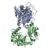
| ||||||||
|---|---|---|---|---|---|---|---|---|---|
| 1 |
| ||||||||
| Unit cell |
| ||||||||
| Components on special symmetry positions |
|
-Components
| #1: Protein | Mass: 36679.246 Da / Num. of mol.: 2 Source method: isolated from a genetically manipulated source Source: (gene. exp.) Mycolicibacterium vanbaalenii (strain DSM 7251 / JCM 13017 / BCRC 16820 / KCTC 9966 / NRRL B-24157 / PYR-1) (bacteria)Gene: Mvan_4516 / Production host: References: UniProt: A1TDP1, Transferases; Transferring nitrogenous groups; Transaminases #2: Water | ChemComp-HOH / |  Mass: 18.015 Da / Num. of mol.: 96 / Source method: isolated from a natural source / Formula: H2O Mass: 18.015 Da / Num. of mol.: 96 / Source method: isolated from a natural source / Formula: H2OHas protein modification | N | |
|---|
-Experimental details
-Experiment
| Experiment | Method: X-RAY DIFFRACTION / Number of used crystals: 1 |
|---|
- Sample preparation
| Crystal | Density Matthews: 2.51 Å3/Da / Density % sol: 50.98 % |
|---|---|
| Crystal grow | Temperature: 298 K / Method: vapor diffusion, hanging drop / Details: 35% Tacsimate pH7.0,0.1 M Bis-Tris propane pH 6.5 |
-Data collection
| Diffraction | Mean temperature: 100 K / Serial crystal experiment: N |
|---|---|
| Diffraction source | Source: SYNCHROTRON / Site: SSRF / Beamline: BL17UM / Wavelength: 1 Å |
| Detector | Type: DECTRIS PILATUS3 2M / Detector: PIXEL / Date: Nov 1, 2021 |
| Radiation | Protocol: SINGLE WAVELENGTH / Monochromatic (M) / Laue (L): M / Scattering type: x-ray |
| Radiation wavelength | Wavelength: 1 Å / Relative weight: 1 |
| Reflection | Resolution: 2.33→30 Å / Num. obs: 34208 / % possible obs: 99.9 % / Redundancy: 40.7 % / CC1/2: 0.996 / CC star: 0.999 / Χ2: 0.059 / Net I/σ(I): 22.2 |
| Reflection shell | Resolution: 2.33→2.41 Å / Redundancy: 38.7 % / Mean I/σ(I) obs: 1.87 / Num. unique obs: 6105 / CC1/2: 0.799 / CC star: 0.942 / Χ2: 0.44 / % possible all: 100 |
- Processing
| Software |
| ||||||||||||||||||||||||||||||||||||||||||||||||||||||||||||||||||||||||||||||||||||||||||||||||||||||||||||||||||||||||||||||||||||||||||||||||||||||||||||||||||||||||||||||||||||||
|---|---|---|---|---|---|---|---|---|---|---|---|---|---|---|---|---|---|---|---|---|---|---|---|---|---|---|---|---|---|---|---|---|---|---|---|---|---|---|---|---|---|---|---|---|---|---|---|---|---|---|---|---|---|---|---|---|---|---|---|---|---|---|---|---|---|---|---|---|---|---|---|---|---|---|---|---|---|---|---|---|---|---|---|---|---|---|---|---|---|---|---|---|---|---|---|---|---|---|---|---|---|---|---|---|---|---|---|---|---|---|---|---|---|---|---|---|---|---|---|---|---|---|---|---|---|---|---|---|---|---|---|---|---|---|---|---|---|---|---|---|---|---|---|---|---|---|---|---|---|---|---|---|---|---|---|---|---|---|---|---|---|---|---|---|---|---|---|---|---|---|---|---|---|---|---|---|---|---|---|---|---|---|---|
| Refinement | Method to determine structure: MOLECULAR REPLACEMENT / Resolution: 2.33→29.08 Å / Cor.coef. Fo:Fc: 0.946 / Cor.coef. Fo:Fc free: 0.902 / SU B: 7.939 / SU ML: 0.186 / Cross valid method: THROUGHOUT / ESU R: 0.281 / ESU R Free: 0.238 / Stereochemistry target values: MAXIMUM LIKELIHOOD / Details: HYDROGENS HAVE BEEN ADDED IN THE RIDING POSITIONS
| ||||||||||||||||||||||||||||||||||||||||||||||||||||||||||||||||||||||||||||||||||||||||||||||||||||||||||||||||||||||||||||||||||||||||||||||||||||||||||||||||||||||||||||||||||||||
| Solvent computation | Ion probe radii: 0.8 Å / Shrinkage radii: 0.8 Å / VDW probe radii: 1.2 Å / Solvent model: MASK | ||||||||||||||||||||||||||||||||||||||||||||||||||||||||||||||||||||||||||||||||||||||||||||||||||||||||||||||||||||||||||||||||||||||||||||||||||||||||||||||||||||||||||||||||||||||
| Displacement parameters | Biso mean: 51.133 Å2
| ||||||||||||||||||||||||||||||||||||||||||||||||||||||||||||||||||||||||||||||||||||||||||||||||||||||||||||||||||||||||||||||||||||||||||||||||||||||||||||||||||||||||||||||||||||||
| Refinement step | Cycle: 1 / Resolution: 2.33→29.08 Å
| ||||||||||||||||||||||||||||||||||||||||||||||||||||||||||||||||||||||||||||||||||||||||||||||||||||||||||||||||||||||||||||||||||||||||||||||||||||||||||||||||||||||||||||||||||||||
| Refine LS restraints |
|