 Movie
Movie Controller

[English] 日本語
 Yorodumi
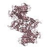
- PDB-8im8: Crystal structure of Periplasmic alpha-amylase (MalS) from E.coli -
+ Open data

- Basic information
| Entry | Database: PDB / ID: 8im8 | ||||||
|---|---|---|---|---|---|---|---|
| Title | Crystal structure of Periplasmic alpha-amylase (MalS) from E.coli | ||||||
 Components Components | Periplasmic alpha-amylase | ||||||
 Keywords Keywords | HYDROLASE / maltooligosaccharides / malS / GH13 / Glycosidase / STRUCTURAL PROTEIN | ||||||
| Function / homology |  Function and homology information Function and homology informationalpha-glucan catabolic process / alpha-amylase / alpha-amylase activity / oligosaccharide catabolic process / outer membrane-bounded periplasmic space / periplasmic space / calcium ion binding Similarity search - Function | ||||||
| Biological species |  | ||||||
| Method |  X-RAY DIFFRACTION / SYNCHROTRON / MOLECULAR REPLACEMENT / Resolution: 2.7 Å X-RAY DIFFRACTION / SYNCHROTRON / MOLECULAR REPLACEMENT / Resolution: 2.7 Å | ||||||
 Authors Authors | An, Y. / Park, J.T. / Park, K.H. / Woo, E.J. | ||||||
| Funding support | 1items
| ||||||
 Citation Citation | Journal: Molecules / Year: 2023 Title: The Distinctive Permutated Domain Structure of Periplasmic alpha-Amylase (MalS) from Glycoside Hydrolase Family 13 Subfamily 19. Authors: An, Y. / Tran, P.L. / Yoo, M.J. / Song, H.N. / Park, K.H. / Kim, T.J. / Park, J.T. / Woo, E.J. | ||||||
| History |
|
- Structure visualization
| Structure viewer | Molecule: MolmilJmol/JSmol |
|---|
- Downloads & links
-Download
| PDBx/mmCIF format | 8im8.cif.gz | 994.4 KB | Display | PDBx/mmCIF format |
|---|---|---|---|---|
| PDB format | pdb8im8.ent.gz | 736 KB | Display | PDB format |
| PDBx/mmJSON format | 8im8.json.gz | Tree view | PDBx/mmJSON format | |
| Others |  Other downloads Other downloads |
-Validation report
| Summary document | 8im8_validation.pdf.gz | 3.2 MB | Display | wwPDB validaton report |
|---|---|---|---|---|
| Full document | 8im8_full_validation.pdf.gz | 3.3 MB | Display | |
| Data in XML | 8im8_validation.xml.gz | 77.8 KB | Display | |
| Data in CIF | 8im8_validation.cif.gz | 107.5 KB | Display | |
| Arichive directory | https://data.pdbj.org/pub/pdb/validation_reports/im/8im8ftp://data.pdbj.org/pub/pdb/validation_reports/im/8im8 | HTTPS FTP |
-Related structure data

-Links
 PDBj
PDBj

- Assembly
| Deposited unit | 
| ||||||||||||
|---|---|---|---|---|---|---|---|---|---|---|---|---|---|
| 1 | 
| ||||||||||||
| 2 | 
| ||||||||||||
| 3 | 
| ||||||||||||
| 4 | 
| ||||||||||||
| Unit cell |
| ||||||||||||
| Components on special symmetry positions |
|
-Components
| #1: Protein | Mass: 75790.352 Da / Num. of mol.: 4 Source method: isolated from a genetically manipulated source Source: (gene. exp.) #2: Chemical | ChemComp-CA /   Mass: 40.078 Da / Num. of mol.: 8 / Source method: obtained synthetically / Formula: Ca / Feature type: SUBJECT OF INVESTIGATION Mass: 40.078 Da / Num. of mol.: 8 / Source method: obtained synthetically / Formula: Ca / Feature type: SUBJECT OF INVESTIGATION#3: Water | ChemComp-HOH / |  Mass: 18.015 Da / Num. of mol.: 292 / Source method: isolated from a natural source / Formula: H2O Mass: 18.015 Da / Num. of mol.: 292 / Source method: isolated from a natural source / Formula: H2OHas ligand of interest | Y | Has protein modification | Y | |
|---|
-Experimental details
-Experiment
| Experiment | Method: X-RAY DIFFRACTION / Number of used crystals: 1 |
|---|
- Sample preparation
| Crystal | Density Matthews: 2.18 Å3/Da / Density % sol: 43.49 % |
|---|---|
| Crystal grow | Temperature: 291.15 K / Method: vapor diffusion, sitting drop Details: 0.1M Sodium citrate tribasic dihydrate pH5.5 20% w/v Polyehtylene glycol 1000 0.1M Lthium sulfate monohydrate |
-Data collection
| Diffraction | Mean temperature: 100 K / Serial crystal experiment: N |
|---|---|
| Diffraction source | Source: SYNCHROTRON / Site: PAL/PLS  / Beamline: 7A (6B, 6C1) / Wavelength: 1 Å / Beamline: 7A (6B, 6C1) / Wavelength: 1 Å |
| Detector | Type: ADSC QUANTUM 270 / Detector: CCD / Date: Sep 30, 2015 / Details: 1EA9, 5A2A |
| Radiation | Protocol: SINGLE WAVELENGTH / Monochromatic (M) / Laue (L): M / Scattering type: x-ray |
| Radiation wavelength | Wavelength: 1 Å / Relative weight: 1 |
| Reflection | Resolution: 2.7→20.02 Å / Num. obs: 69932 / % possible obs: 99.83 % / Redundancy: 11.4 % / Biso Wilson estimate: 44.25 Å2 / Rrim(I) all: 0.123 / Net I/σ(I): 168 |
| Reflection shell | Resolution: 2.7→2.77 Å / Mean I/σ(I) obs: 5.5 / Num. unique obs: 4802 / Rrim(I) all: 0.684 / % possible all: 98.5 |
- Processing
| Software |
| |||||||||||||||||||||||||||||||||||||||||||||||||||||||||||||||||||||||||||||||||||||||||||||||||||||||||
|---|---|---|---|---|---|---|---|---|---|---|---|---|---|---|---|---|---|---|---|---|---|---|---|---|---|---|---|---|---|---|---|---|---|---|---|---|---|---|---|---|---|---|---|---|---|---|---|---|---|---|---|---|---|---|---|---|---|---|---|---|---|---|---|---|---|---|---|---|---|---|---|---|---|---|---|---|---|---|---|---|---|---|---|---|---|---|---|---|---|---|---|---|---|---|---|---|---|---|---|---|---|---|---|---|---|---|
| Refinement | Method to determine structure: MOLECULAR REPLACEMENT Starting model: 1EA9, 5A2A Resolution: 2.7→20.02 Å / SU ML: 0.3187 / Cross valid method: FREE R-VALUE / σ(F): 1.97 / Phase error: 26.7265 Stereochemistry target values: GeoStd + Monomer Library + CDL v1.2
| |||||||||||||||||||||||||||||||||||||||||||||||||||||||||||||||||||||||||||||||||||||||||||||||||||||||||
| Solvent computation | Shrinkage radii: 0.9 Å / VDW probe radii: 1.11 Å / Solvent model: FLAT BULK SOLVENT MODEL | |||||||||||||||||||||||||||||||||||||||||||||||||||||||||||||||||||||||||||||||||||||||||||||||||||||||||
| Displacement parameters | Biso mean: 48.44 Å2 | |||||||||||||||||||||||||||||||||||||||||||||||||||||||||||||||||||||||||||||||||||||||||||||||||||||||||
| Refinement step | Cycle: LAST / Resolution: 2.7→20.02 Å
| |||||||||||||||||||||||||||||||||||||||||||||||||||||||||||||||||||||||||||||||||||||||||||||||||||||||||
| Refine LS restraints |
| |||||||||||||||||||||||||||||||||||||||||||||||||||||||||||||||||||||||||||||||||||||||||||||||||||||||||
| LS refinement shell |
| |||||||||||||||||||||||||||||||||||||||||||||||||||||||||||||||||||||||||||||||||||||||||||||||||||||||||
| Refinement TLS params. | Method: refined / Origin x: -13.3721715185 Å / Origin y: 42.4147738286 Å / Origin z: 340.955066158 Å
| |||||||||||||||||||||||||||||||||||||||||||||||||||||||||||||||||||||||||||||||||||||||||||||||||||||||||
| Refinement TLS group | Selection details: all |
