 Movie
Movie Controller

[English] 日本語
 Yorodumi
- PDB-8ila: Crystal structure of LmbT from Streptomyces lincolnensis NRRL ISP... -
+ Open data

- Basic information
| Entry | Database: PDB / ID: 8ila | |||||||||
|---|---|---|---|---|---|---|---|---|---|---|
| Title | Crystal structure of LmbT from Streptomyces lincolnensis NRRL ISP-5355 in complex with substrates | |||||||||
 Components Components | Glycosyltransferase | |||||||||
 Keywords Keywords | BIOSYNTHETIC PROTEIN / Glycosyltransferase | |||||||||
| Function / homology | Glycosyl transferase, family 1 / Glycosyl transferases group 1 / glycosyltransferase activity / GUANOSINE-5'-DIPHOSPHATE / Chem-Q3L / D-inositol 3-phosphate glycosyltransferase Function and homology information Function and homology information | |||||||||
| Biological species |  Streptomyces lincolnensis (bacteria) Streptomyces lincolnensis (bacteria) | |||||||||
| Method |  X-RAY DIFFRACTION / SYNCHROTRON / MOLECULAR REPLACEMENT / Resolution: 2.79 Å X-RAY DIFFRACTION / SYNCHROTRON / MOLECULAR REPLACEMENT / Resolution: 2.79 Å | |||||||||
 Authors Authors | Dai, Y. / Qiao, H. / Xia, M. / Fang, P. / Liu, W. | |||||||||
| Funding support |  China, 2items China, 2items
| |||||||||
 Citation Citation | Journal: Acs Chem.Biol. / Year: 2023 Title: Structural Basis of Low-Molecular-Weight Thiol Glycosylation in Lincomycin A Biosynthesis. Authors: Dai, Y. / Cheng, Y. / Ding, W. / Qiao, H. / Zhang, D. / Zhong, G. / Xia, M. / Tao, J. / Sun, P. / Fang, P. / Liu, W. | |||||||||
| History |
|
- Structure visualization
| Structure viewer | Molecule: MolmilJmol/JSmol |
|---|
- Downloads & links
-Download
| PDBx/mmCIF format | 8ila.cif.gz | 647.7 KB | Display | PDBx/mmCIF format |
|---|---|---|---|---|
| PDB format | pdb8ila.ent.gz | 537.8 KB | Display | PDB format |
| PDBx/mmJSON format | 8ila.json.gz | Tree view | PDBx/mmJSON format | |
| Others |  Other downloads Other downloads |
-Validation report
| Summary document | 8ila_validation.pdf.gz | 2.6 MB | Display | wwPDB validaton report |
|---|---|---|---|---|
| Full document | 8ila_full_validation.pdf.gz | 2.7 MB | Display | |
| Data in XML | 8ila_validation.xml.gz | 64.8 KB | Display | |
| Data in CIF | 8ila_validation.cif.gz | 86 KB | Display | |
| Arichive directory | https://data.pdbj.org/pub/pdb/validation_reports/il/8ilaftp://data.pdbj.org/pub/pdb/validation_reports/il/8ila | HTTPS FTP |
-Related structure data

-Links
 PDBj
PDBj
- Assembly
| Deposited unit | 
| ||||||||
|---|---|---|---|---|---|---|---|---|---|
| 1 |
| ||||||||
| Unit cell |
|
-Components
| #1: Protein | Mass: 49408.723 Da / Num. of mol.: 4 Source method: isolated from a genetically manipulated source Source: (gene. exp.) Streptomyces lincolnensis (bacteria) / Gene: GJU35_01490 / Production host: #2: Chemical | ChemComp-GDP /   Type: RNA linking / Mass: 443.201 Da / Num. of mol.: 4 / Source method: obtained synthetically / Formula: C10H15N5O11P2 / Comment: GDP, energy-carrying molecule*YM Type: RNA linking / Mass: 443.201 Da / Num. of mol.: 4 / Source method: obtained synthetically / Formula: C10H15N5O11P2 / Comment: GDP, energy-carrying molecule*YM#3: Chemical | ChemComp-Q3L / ( 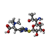  Mass: 435.516 Da / Num. of mol.: 4 / Source method: obtained synthetically / Formula: C17H31N4O7S / Feature type: SUBJECT OF INVESTIGATION Mass: 435.516 Da / Num. of mol.: 4 / Source method: obtained synthetically / Formula: C17H31N4O7S / Feature type: SUBJECT OF INVESTIGATION#4: Water | ChemComp-HOH / |  Mass: 18.015 Da / Num. of mol.: 57 / Source method: isolated from a natural source / Formula: H2O Mass: 18.015 Da / Num. of mol.: 57 / Source method: isolated from a natural source / Formula: H2OHas ligand of interest | Y | |
|---|
-Experimental details
-Experiment
| Experiment | Method: X-RAY DIFFRACTION / Number of used crystals: 1 |
|---|
- Sample preparation
| Crystal | Density Matthews: 3.12 Å3/Da / Density % sol: 60.55 % |
|---|---|
| Crystal grow | Temperature: 291 K / Method: vapor diffusion, sitting drop Details: 8% PEG 20000, 0.05 M Tris pH 8.5, 1.2 M Sodium formate |
-Data collection
| Diffraction | Mean temperature: 100 K / Serial crystal experiment: N |
|---|---|
| Diffraction source | Source: SYNCHROTRON / Site: SSRF / Beamline: BL10U2 / Wavelength: 0.979183 Å |
| Detector | Type: DECTRIS EIGER X 16M / Detector: PIXEL / Date: Oct 8, 2022 |
| Radiation | Protocol: SINGLE WAVELENGTH / Monochromatic (M) / Laue (L): M / Scattering type: x-ray |
| Radiation wavelength | Wavelength: 0.979183 Å / Relative weight: 1 |
| Reflection | Resolution: 2.79→100.22 Å / Num. obs: 62181 / % possible obs: 99.7 % / Redundancy: 7.1 % / CC1/2: 0.998 / Rmerge(I) obs: 0.091 / Net I/σ(I): 16.2 |
| Reflection shell | Resolution: 2.79→2.94 Å / Num. unique obs: 9020 / CC1/2: 0.828 / % possible all: 100 |
- Processing
| Software |
| |||||||||||||||||||||||||||||||||||||||||||||||||||||||||||||||||||||||||||||||||||||||||||||||||||||||||||||||||||||||||||||||||||||||||||||||||||||||||||||||||
|---|---|---|---|---|---|---|---|---|---|---|---|---|---|---|---|---|---|---|---|---|---|---|---|---|---|---|---|---|---|---|---|---|---|---|---|---|---|---|---|---|---|---|---|---|---|---|---|---|---|---|---|---|---|---|---|---|---|---|---|---|---|---|---|---|---|---|---|---|---|---|---|---|---|---|---|---|---|---|---|---|---|---|---|---|---|---|---|---|---|---|---|---|---|---|---|---|---|---|---|---|---|---|---|---|---|---|---|---|---|---|---|---|---|---|---|---|---|---|---|---|---|---|---|---|---|---|---|---|---|---|---|---|---|---|---|---|---|---|---|---|---|---|---|---|---|---|---|---|---|---|---|---|---|---|---|---|---|---|---|---|---|---|
| Refinement | Method to determine structure: MOLECULAR REPLACEMENT / Resolution: 2.79→68.87 Å / SU ML: 0.39 / Cross valid method: FREE R-VALUE / σ(F): 1.33 / Phase error: 30.92 / Stereochemistry target values: ML
| |||||||||||||||||||||||||||||||||||||||||||||||||||||||||||||||||||||||||||||||||||||||||||||||||||||||||||||||||||||||||||||||||||||||||||||||||||||||||||||||||
| Solvent computation | Shrinkage radii: 0.9 Å / VDW probe radii: 1.1 Å / Solvent model: FLAT BULK SOLVENT MODEL | |||||||||||||||||||||||||||||||||||||||||||||||||||||||||||||||||||||||||||||||||||||||||||||||||||||||||||||||||||||||||||||||||||||||||||||||||||||||||||||||||
| Refinement step | Cycle: LAST / Resolution: 2.79→68.87 Å
| |||||||||||||||||||||||||||||||||||||||||||||||||||||||||||||||||||||||||||||||||||||||||||||||||||||||||||||||||||||||||||||||||||||||||||||||||||||||||||||||||
| Refine LS restraints |
| |||||||||||||||||||||||||||||||||||||||||||||||||||||||||||||||||||||||||||||||||||||||||||||||||||||||||||||||||||||||||||||||||||||||||||||||||||||||||||||||||
| LS refinement shell |
| |||||||||||||||||||||||||||||||||||||||||||||||||||||||||||||||||||||||||||||||||||||||||||||||||||||||||||||||||||||||||||||||||||||||||||||||||||||||||||||||||
| Refinement TLS params. | Method: refined / Origin x: 39.3314 Å / Origin y: 20.9171 Å / Origin z: 31.341 Å
| |||||||||||||||||||||||||||||||||||||||||||||||||||||||||||||||||||||||||||||||||||||||||||||||||||||||||||||||||||||||||||||||||||||||||||||||||||||||||||||||||
| Refinement TLS group | Selection details: all |
