 Movie
Movie Controller

[English] 日本語
 Yorodumi
- PDB-8ika: Cryo-EM structure of the encapsulin shell from Mycobacterium tube... -
+ Open data

- Basic information
| Entry | Database: PDB / ID: 8ika | ||||||||||||
|---|---|---|---|---|---|---|---|---|---|---|---|---|---|
| Title | Cryo-EM structure of the encapsulin shell from Mycobacterium tuberculosis | ||||||||||||
 Components Components | Type 1 encapsulin shell protein | ||||||||||||
 Keywords Keywords | VIRUS LIKE PARTICLE / CFP-29 / Mycobacterium tuberculosis / cryo-EM / encapsulin | ||||||||||||
| Function / homology | Type 1 encapsulin shell protein / Encapsulating protein for peroxidase / : / encapsulin nanocompartment / extracellular region / plasma membrane / Type 1 encapsulin shell protein Function and homology information Function and homology information | ||||||||||||
| Biological species |   Mycobacterium tuberculosis (bacteria) Mycobacterium tuberculosis (bacteria) | ||||||||||||
| Method | ELECTRON MICROSCOPY / single particle reconstruction / cryo EM / Resolution: 2.75 Å | ||||||||||||
 Authors Authors | Du, Z. / Lai, Y. / Gao, Y. / Rao, Z. / Gong, H. | ||||||||||||
| Funding support |  China, 3items China, 3items
| ||||||||||||
 Citation Citation | Journal: To Be Published Title: Structure of mycobacterium tuberculosis encapsulin Authors: Du, Z. / Lai, Y. / Gao, Y. / Rao, Z. / Gong, H. | ||||||||||||
| History |
|
- Structure visualization
| Structure viewer | Molecule: MolmilJmol/JSmol |
|---|
- Downloads & links
-Download
| PDBx/mmCIF format | 8ika.cif.gz | 2.5 MB | Display | PDBx/mmCIF format |
|---|---|---|---|---|
| PDB format | pdb8ika.ent.gz | Display | PDB format | |
| PDBx/mmJSON format | 8ika.json.gz | Tree view | PDBx/mmJSON format | |
| Others |  Other downloads Other downloads |
-Validation report
| Summary document | 8ika_validation.pdf.gz | 380.5 KB | Display | wwPDB validaton report |
|---|---|---|---|---|
| Full document | 8ika_full_validation.pdf.gz | 391.7 KB | Display | |
| Data in XML | 8ika_validation.xml.gz | 257.6 KB | Display | |
| Data in CIF | 8ika_validation.cif.gz | 309.8 KB | Display | |
| Arichive directory | https://data.pdbj.org/pub/pdb/validation_reports/ik/8ikaftp://data.pdbj.org/pub/pdb/validation_reports/ik/8ika | HTTPS FTP |
-Related structure data
| Related structure data | 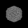 35507MC M: map data used to model this data C: citing same article ( |
|---|---|
| Similar structure data |
-Links
 PDBj
PDBj
- Assembly
| Deposited unit | 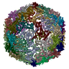
|
|---|---|
| 1 |
|
-Components
| #1: Protein | Mass: 28863.346 Da / Num. of mol.: 60 Source method: isolated from a genetically manipulated source Source: (gene. exp.) Mycobacterium tuberculosis (strain ATCC 25618 / H37Rv) (bacteria)Gene: enc, cfp29, Rv0798c / Production host: |
|---|
-Experimental details
-Experiment
| Experiment | Method: ELECTRON MICROSCOPY |
|---|---|
| EM experiment | Aggregation state: PARTICLE / 3D reconstruction method: single particle reconstruction |
- Sample preparation
| Component | Name: Mycobacterium tuberculosis encapsulin / Type: COMPLEX / Entity ID: all / Source: MULTIPLE SOURCES |
|---|---|
| Molecular weight | Value: 2 MDa / Experimental value: YES |
| Source (natural) | Organism: Mycobacterium tuberculosis (bacteria) |
| Source (recombinant) | Organism: |
| Buffer solution | pH: 7.4 |
| Specimen | Embedding applied: NO / Shadowing applied: NO / Staining applied: NO / Vitrification applied: YES |
| Vitrification | Cryogen name: ETHANE |
- Electron microscopy imaging
| Experimental equipment |  Model: Titan Krios / Image courtesy: FEI Company |
|---|---|
| Microscopy | Model: FEI TITAN KRIOS |
| Electron gun | Electron source:  FIELD EMISSION GUN / Accelerating voltage: 300 kV / Illumination mode: FLOOD BEAM FIELD EMISSION GUN / Accelerating voltage: 300 kV / Illumination mode: FLOOD BEAM |
| Electron lens | Mode: BRIGHT FIELD / Nominal defocus max: 2400 nm / Nominal defocus min: 1200 nm |
| Image recording | Electron dose: 60 e/Å2 / Film or detector model: GATAN K3 BIOQUANTUM (6k x 4k) |
- Processing
| CTF correction | Type: PHASE FLIPPING AND AMPLITUDE CORRECTION |
|---|---|
| 3D reconstruction | Resolution: 2.75 Å / Resolution method: FSC 0.143 CUT-OFF / Num. of particles: 251715 / Symmetry type: POINT |
