 Movie
Movie Controller

[English] 日本語
 Yorodumi
- PDB-8iib: Crystal structure of Israeli acute paralysis virus RNA-dependent ... -
+ Open data

- Basic information
| Entry | Database: PDB / ID: 8iib | |||||||||
|---|---|---|---|---|---|---|---|---|---|---|
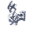
| Title | Crystal structure of Israeli acute paralysis virus RNA-dependent RNA polymerase delta85 mutant (residues 86-546) | |||||||||
 Components Components | Polymerase polyprotein | |||||||||
 Keywords Keywords | TRANSFERASE / RNA-dependent RNA polymerase | |||||||||
| Function / homology | :  Function and homology information Function and homology information | |||||||||
| Biological species |  Israeli acute paralysis virus Israeli acute paralysis virus | |||||||||
| Method |  X-RAY DIFFRACTION / SYNCHROTRON / SAD / Resolution: 2.69 Å X-RAY DIFFRACTION / SYNCHROTRON / SAD / Resolution: 2.69 Å | |||||||||
 Authors Authors | Fang, X. / Lu, G. / Hou, C. / Gong, P. | |||||||||
| Funding support |  China, 2items China, 2items
| |||||||||
 Citation Citation | Journal: Virol Sin / Year: 2023 Title: Unusual substructure conformations observed in crystal structures of a dicistrovirus RNA-dependent RNA polymerase suggest contribution of the N-terminal extension in proper folding. Authors: Fang, X. / Lu, G. / Deng, Y. / Yang, S. / Hou, C. / Gong, P. | |||||||||
| History |
|
- Structure visualization
| Structure viewer | Molecule: MolmilJmol/JSmol |
|---|
- Downloads & links
-Download
| PDBx/mmCIF format | 8iib.cif.gz | 157.6 KB | Display | PDBx/mmCIF format |
|---|---|---|---|---|
| PDB format | pdb8iib.ent.gz | 117.9 KB | Display | PDB format |
| PDBx/mmJSON format | 8iib.json.gz | Tree view | PDBx/mmJSON format | |
| Others |  Other downloads Other downloads |
-Validation report
| Arichive directory | https://data.pdbj.org/pub/pdb/validation_reports/ii/8iibftp://data.pdbj.org/pub/pdb/validation_reports/ii/8iib | HTTPS FTP |
|---|
-Related structure data
-Links
 PDBj
PDBj- Assembly
| Deposited unit | 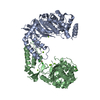
| ||||||||
|---|---|---|---|---|---|---|---|---|---|
| 1 | 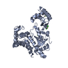
| ||||||||
| 2 | 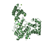
| ||||||||
| Unit cell |
| ||||||||
| Components on special symmetry positions |
|
-Components
| #1: Protein | Mass: 54888.414 Da / Num. of mol.: 2 / Mutation: N510D Source method: isolated from a genetically manipulated source Details: GB:MG599488.1 / Source: (gene. exp.) Israeli acute paralysis virus / Production host:  #2: Chemical | ChemComp-CD /   Mass: 112.411 Da / Num. of mol.: 4 / Source method: obtained synthetically / Formula: Cd / Feature type: SUBJECT OF INVESTIGATION Mass: 112.411 Da / Num. of mol.: 4 / Source method: obtained synthetically / Formula: Cd / Feature type: SUBJECT OF INVESTIGATION#3: Chemical | ChemComp-MG /   Mass: 24.305 Da / Num. of mol.: 8 / Source method: obtained synthetically / Formula: Mg Mass: 24.305 Da / Num. of mol.: 8 / Source method: obtained synthetically / Formula: Mg#4: Chemical | ChemComp-CL /   Mass: 35.453 Da / Num. of mol.: 7 / Source method: obtained synthetically / Formula: Cl / Feature type: SUBJECT OF INVESTIGATION Mass: 35.453 Da / Num. of mol.: 7 / Source method: obtained synthetically / Formula: Cl / Feature type: SUBJECT OF INVESTIGATION#5: Water | ChemComp-HOH / |  Mass: 18.015 Da / Num. of mol.: 160 / Source method: isolated from a natural source / Formula: H2O Mass: 18.015 Da / Num. of mol.: 160 / Source method: isolated from a natural source / Formula: H2OHas ligand of interest | Y | |
|---|
-Experimental details
-Experiment
| Experiment | Method: X-RAY DIFFRACTION / Number of used crystals: 1 |
|---|
- Sample preparation
| Crystal | Density Matthews: 3.12 Å3/Da / Density % sol: 60.53 % |
|---|---|
| Crystal grow | Temperature: 289 K / Method: evaporation / pH: 8.5 / Details: PEG 4000 |
-Data collection
| Diffraction | Mean temperature: 100 K / Serial crystal experiment: N | |||||||||||||||||||||||||||||||||||||||||||||||||||||||||||||||||||||||||||||||||||||||||||||||||||
|---|---|---|---|---|---|---|---|---|---|---|---|---|---|---|---|---|---|---|---|---|---|---|---|---|---|---|---|---|---|---|---|---|---|---|---|---|---|---|---|---|---|---|---|---|---|---|---|---|---|---|---|---|---|---|---|---|---|---|---|---|---|---|---|---|---|---|---|---|---|---|---|---|---|---|---|---|---|---|---|---|---|---|---|---|---|---|---|---|---|---|---|---|---|---|---|---|---|---|---|---|
| Diffraction source | Source: SYNCHROTRON / Site: SSRF / Beamline: BL17U1 / Wavelength: 0.9792 Å | |||||||||||||||||||||||||||||||||||||||||||||||||||||||||||||||||||||||||||||||||||||||||||||||||||
| Detector | Type: DECTRIS EIGER X 16M / Detector: PIXEL / Date: Dec 6, 2018 | |||||||||||||||||||||||||||||||||||||||||||||||||||||||||||||||||||||||||||||||||||||||||||||||||||
| Radiation | Protocol: SINGLE WAVELENGTH / Monochromatic (M) / Laue (L): M / Scattering type: x-ray | |||||||||||||||||||||||||||||||||||||||||||||||||||||||||||||||||||||||||||||||||||||||||||||||||||
| Radiation wavelength | Wavelength: 0.9792 Å / Relative weight: 1 | |||||||||||||||||||||||||||||||||||||||||||||||||||||||||||||||||||||||||||||||||||||||||||||||||||
| Reflection | Resolution: 2.69→23.8 Å / Num. obs: 38083 / % possible obs: 100 % / Redundancy: 10.1 % / Rmerge(I) obs: 0.041 / Rpim(I) all: 0.014 / Rrim(I) all: 0.043 / Χ2: 0.922 / Net I/σ(I): 16.2 | |||||||||||||||||||||||||||||||||||||||||||||||||||||||||||||||||||||||||||||||||||||||||||||||||||
| Reflection shell | Diffraction-ID: 1
|
- Processing
| Software |
| ||||||||||||||||||||||||||||||||||||||||||||||||||||||||||||||||||||||||||||||||||||||||||||||||||
|---|---|---|---|---|---|---|---|---|---|---|---|---|---|---|---|---|---|---|---|---|---|---|---|---|---|---|---|---|---|---|---|---|---|---|---|---|---|---|---|---|---|---|---|---|---|---|---|---|---|---|---|---|---|---|---|---|---|---|---|---|---|---|---|---|---|---|---|---|---|---|---|---|---|---|---|---|---|---|---|---|---|---|---|---|---|---|---|---|---|---|---|---|---|---|---|---|---|---|---|
| Refinement | Method to determine structure: SAD / Resolution: 2.69→23.41 Å / SU ML: 0.31 / Cross valid method: THROUGHOUT / σ(F): 1.37 / Phase error: 25.13 / Stereochemistry target values: ML
| ||||||||||||||||||||||||||||||||||||||||||||||||||||||||||||||||||||||||||||||||||||||||||||||||||
| Solvent computation | Shrinkage radii: 0.9 Å / VDW probe radii: 1.11 Å / Solvent model: FLAT BULK SOLVENT MODEL | ||||||||||||||||||||||||||||||||||||||||||||||||||||||||||||||||||||||||||||||||||||||||||||||||||
| Displacement parameters | Biso max: 94.46 Å2 / Biso mean: 40.2077 Å2 / Biso min: 10.8 Å2 | ||||||||||||||||||||||||||||||||||||||||||||||||||||||||||||||||||||||||||||||||||||||||||||||||||
| Refinement step | Cycle: final / Resolution: 2.69→23.41 Å
| ||||||||||||||||||||||||||||||||||||||||||||||||||||||||||||||||||||||||||||||||||||||||||||||||||
| LS refinement shell | Refine-ID: X-RAY DIFFRACTION / Rfactor Rfree error: 0 / Total num. of bins used: 13
|
