 Movie
Movie Controller

[English] 日本語
 Yorodumi
- PDB-8iia: Crystal structure of the oligomeric state of the extracellular do... -
+ Open data

- Basic information
| Entry | Database: PDB / ID: 8iia | ||||||||||||||||||
|---|---|---|---|---|---|---|---|---|---|---|---|---|---|---|---|---|---|---|---|
| Title | Crystal structure of the oligomeric state of the extracellular domain of human myelin protein zero(MPZ/P0) | ||||||||||||||||||
 Components Components | Myelin protein P0 | ||||||||||||||||||
 Keywords Keywords | CELL ADHESION / membrane adhesion / oligomeric state | ||||||||||||||||||
| Function / homology |  Function and homology information Function and homology informationcell aggregation / : / EGR2 and SOX10-mediated initiation of Schwann cell myelination / myelination / myelin sheath / chemical synaptic transmission / synapse / structural molecule activity / plasma membrane Similarity search - Function | ||||||||||||||||||
| Biological species |  Homo sapiens (human) Homo sapiens (human) | ||||||||||||||||||
| Method |  X-RAY DIFFRACTION / SYNCHROTRON / MOLECULAR REPLACEMENT / Resolution: 2.09 Å X-RAY DIFFRACTION / SYNCHROTRON / MOLECULAR REPLACEMENT / Resolution: 2.09 Å | ||||||||||||||||||
 Authors Authors | Sakakura, M. / Tanabe, M. / Mio, K. | ||||||||||||||||||
| Funding support |  Japan, 5items Japan, 5items
| ||||||||||||||||||
 Citation Citation | Journal: Structure / Year: 2023 Title: Structural bases for the Charcot-Marie-Tooth disease induced by single amino acid substitutions of myelin protein zero. Authors: Sakakura, M. / Tanabe, M. / Mori, M. / Takahashi, H. / Mio, K. | ||||||||||||||||||
| History |
|
- Structure visualization
| Structure viewer | Molecule: MolmilJmol/JSmol |
|---|
- Downloads & links
-Download
| PDBx/mmCIF format | 8iia.cif.gz | 47.2 KB | Display | PDBx/mmCIF format |
|---|---|---|---|---|
| PDB format | pdb8iia.ent.gz | 26.2 KB | Display | PDB format |
| PDBx/mmJSON format | 8iia.json.gz | Tree view | PDBx/mmJSON format | |
| Others |  Other downloads Other downloads |
-Validation report
| Arichive directory | https://data.pdbj.org/pub/pdb/validation_reports/ii/8iiaftp://data.pdbj.org/pub/pdb/validation_reports/ii/8iia | HTTPS FTP |
|---|
-Related structure data
| Related structure data | 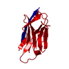 1neuS S: Starting model for refinement |
|---|---|
| Similar structure data |
-Links
 PDBj
PDBj


- Assembly
| Deposited unit | 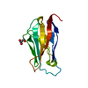
| ||||||||||||
|---|---|---|---|---|---|---|---|---|---|---|---|---|---|
| 1 |
| ||||||||||||
| Unit cell |
|
-Components
| #1: Protein | Mass: 14066.645 Da / Num. of mol.: 1 / Fragment: extracellular domain Source method: isolated from a genetically manipulated source Source: (gene. exp.) Homo sapiens (human) / Gene: MPZ / Production host:  |
|---|---|
| #2: Chemical | ChemComp-GOL /   Mass: 92.094 Da / Num. of mol.: 1 / Source method: obtained synthetically / Formula: C3H8O3 Mass: 92.094 Da / Num. of mol.: 1 / Source method: obtained synthetically / Formula: C3H8O3 |
| #3: Water | ChemComp-HOH /  Mass: 18.015 Da / Num. of mol.: 51 / Source method: isolated from a natural source / Formula: H2O Mass: 18.015 Da / Num. of mol.: 51 / Source method: isolated from a natural source / Formula: H2O |
| Has ligand of interest | N |
| Has protein modification | Y |
-Experimental details
-Experiment
| Experiment | Method: X-RAY DIFFRACTION / Number of used crystals: 1 |
|---|
- Sample preparation
| Crystal | Density Matthews: 3.16 Å3/Da / Density % sol: 61.1 % |
|---|---|
| Crystal grow | Temperature: 293 K / Method: vapor diffusion, sitting drop / pH: 8.5 / Details: 100 mM Tris-HCl, 300 mM magnesium-formate |
-Data collection
| Diffraction | Mean temperature: 100 K / Serial crystal experiment: N |
|---|---|
| Diffraction source | Source: SYNCHROTRON / Site: SPring-8 / Beamline: BL32XU / Wavelength: 1 Å |
| Detector | Type: DECTRIS EIGER X 9M / Detector: PIXEL / Date: Apr 1, 2020 |
| Radiation | Protocol: SINGLE WAVELENGTH / Monochromatic (M) / Laue (L): M / Scattering type: x-ray |
| Radiation wavelength | Wavelength: 1 Å / Relative weight: 1 |
| Reflection | Resolution: 2.09→44.49 Å / Num. obs: 10861 / % possible obs: 100 % / Redundancy: 26.8 % / Biso Wilson estimate: 49.75 Å2 / CC1/2: 0.998 / Rmerge(I) obs: 0.141 / Rpim(I) all: 0.028 / Net I/av σ(I): 13.9 / Net I/σ(I): 13.9 |
| Reflection shell | Resolution: 2.09→2.14 Å / Redundancy: 27.4 % / Rmerge(I) obs: 3.297 / Mean I/σ(I) obs: 1.1 / Num. unique obs: 815 / CC1/2: 0.815 / Rpim(I) all: 0.634 / % possible all: 99.5 |
- Processing
| Software |
| |||||||||||||||||||||||||||||||||||
|---|---|---|---|---|---|---|---|---|---|---|---|---|---|---|---|---|---|---|---|---|---|---|---|---|---|---|---|---|---|---|---|---|---|---|---|---|
| Refinement | Method to determine structure: MOLECULAR REPLACEMENT Starting model: 1NEU Resolution: 2.09→36.27 Å / SU ML: 0.2521 / Cross valid method: FREE R-VALUE / σ(F): 1.33 / Phase error: 30.1569 Stereochemistry target values: GeoStd + Monomer Library + CDL v1.2
| |||||||||||||||||||||||||||||||||||
| Solvent computation | Shrinkage radii: 0.9 Å / VDW probe radii: 1.11 Å / Solvent model: FLAT BULK SOLVENT MODEL | |||||||||||||||||||||||||||||||||||
| Displacement parameters | Biso mean: 52.75 Å2 | |||||||||||||||||||||||||||||||||||
| Refinement step | Cycle: LAST / Resolution: 2.09→36.27 Å
| |||||||||||||||||||||||||||||||||||
| Refine LS restraints |
| |||||||||||||||||||||||||||||||||||
| LS refinement shell |
|
