| 登録情報 | データベース: PDB / ID: 8iey
|
|---|
| タイトル | Aquifex aeolicus TsaD-TsaB |
|---|
 要素 要素 | - Gcp-like domain-containing protein
- tRNA N6-adenosine threonylcarbamoyltransferase
|
|---|
 キーワード キーワード | TRANSFERASE / tRNA t6A-modifying enzyme / TsaD-TsaB complex |
|---|
| 機能・相同性 |  機能・相同性情報 機能・相同性情報
N6-L-threonylcarbamoyladenine synthase / tRNA N(6)-L-threonylcarbamoyladenine synthase activity / tRNA threonylcarbamoyladenosine modification / iron ion binding / cytosol / cytoplasm類似検索 - 分子機能 tRNA N6-adenosine threonylcarbamoyltransferase, TsaD / Peptidase M22, conserved site / Glycoprotease family signature. / Kae1/TsaD family / Gcp-like domain / tRNA N6-adenosine threonylcarbamoyltransferase / ATPase, nucleotide binding domain類似検索 - ドメイン・相同性 : / DI(HYDROXYETHYL)ETHER / Gcp-like domain-containing protein / tRNA N6-adenosine threonylcarbamoyltransferase類似検索 - 構成要素 |
|---|
| 生物種 |   Aquifex aeolicus (バクテリア) Aquifex aeolicus (バクテリア) |
|---|
| 手法 |  X線回折 / 分子置換 / 解像度: 2 Å X線回折 / 分子置換 / 解像度: 2 Å |
|---|
 データ登録者 データ登録者 | Lu, S.Z. / Zhang, W.H. |
|---|
| 資金援助 |  中国, 1件 中国, 1件 | 組織 | 認可番号 | 国 |
|---|
| National Natural Science Foundation of China (NSFC) | 32000847 | 中国 |
|
|---|
 引用 引用 | ジャーナル: J.Biol.Chem. / 年: 2024
タイトル: Structure-function analysis of tRNA t 6 A-catalysis, assembly, and thermostability of Aquifex aeolicus TsaD 2 B 2 tetramer in complex with TsaE.
著者: Lu, S. / Jin, M. / Yu, Z. / Zhang, W. |
|---|
| 履歴 | | 登録 | 2023年2月16日 | 登録サイト: PDBJ / 処理サイト: PDBJ |
|---|
| 改定 1.0 | 2024年3月20日 | Provider: repository / タイプ: Initial release |
|---|
| 改定 1.1 | 2024年11月6日 | Group: Structure summary
カテゴリ: pdbx_entry_details / pdbx_modification_feature
Item: _pdbx_entry_details.has_protein_modification |
|---|
| 改定 1.2 | 2024年11月27日 | Group: Database references / カテゴリ: citation / citation_author
Item: _citation.country / _citation.journal_abbrev ..._citation.country / _citation.journal_abbrev / _citation.journal_id_ASTM / _citation.journal_id_CSD / _citation.journal_id_ISSN / _citation.page_first / _citation.page_last / _citation.pdbx_database_id_DOI / _citation.pdbx_database_id_PubMed / _citation.title / _citation.year |
|---|
| 改定 1.3 | 2024年12月11日 | Group: Database references / カテゴリ: citation / Item: _citation.journal_volume / _citation.title |
|---|
|
|---|
 ムービー
ムービー コントローラー


 データを開く
 基本情報
基本情報 構造の表示
構造の表示 ダウンロードとリンク
 その他のダウンロード
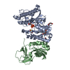
 PDBj
PDBj 集合体
集合体


 分子量: 55.845 Da / 分子数: 1 / 由来タイプ: 合成 / 式: Fe / タイプ: SUBJECT OF INVESTIGATION
 分子量: 106.120 Da / 分子数: 5 / 由来タイプ: 天然 / 式: C4H10O3
 分子量: 18.015 Da / 分子数: 238 / 由来タイプ: 天然 / 式: H2O
 試料調製
試料調製 解析
解析