 Movie
Movie Controller

[English] 日本語
 Yorodumi
- PDB-8idw: Crystal structure of Fic protein from Mycoplasma mycoides in comp... -
+ Open data

- Basic information
| Entry | Database: PDB / ID: 8idw | ||||||
|---|---|---|---|---|---|---|---|
| Title | Crystal structure of Fic protein from Mycoplasma mycoides in complex with AMPPNP | ||||||
 Components Components | Cell filamentation protein Fic | ||||||
 Keywords Keywords | TRANSFERASE / Fic protein / AMP transferase | ||||||
| Function / homology | Fido-like domain superfamily / protein adenylyltransferase / Fic/DOC family / Fido domain / Fido domain profile. / transferase activity / AMP PHOSPHORAMIDATE / protein adenylyltransferase Function and homology information Function and homology information | ||||||
| Biological species |  Mycoplasma mycoides subsp. capri (bacteria) Mycoplasma mycoides subsp. capri (bacteria) | ||||||
| Method |  X-RAY DIFFRACTION / SYNCHROTRON / MOLECULAR REPLACEMENT / Resolution: 2.304 Å X-RAY DIFFRACTION / SYNCHROTRON / MOLECULAR REPLACEMENT / Resolution: 2.304 Å | ||||||
 Authors Authors | Zhang, H. / Zhao, H.F. | ||||||
| Funding support |  China, 1items China, 1items
| ||||||
 Citation Citation | Journal: To Be Published Title: Crystal structure of Fic protein from Mycoplasma mycoides in complex with AMPPNP Authors: Zhang, H. / Zhao, H.F. | ||||||
| History |
|
- Structure visualization
| Structure viewer | Molecule: MolmilJmol/JSmol |
|---|
- Downloads & links
-Download
| PDBx/mmCIF format | 8idw.cif.gz | 167.8 KB | Display | PDBx/mmCIF format |
|---|---|---|---|---|
| PDB format | pdb8idw.ent.gz | 133.6 KB | Display | PDB format |
| PDBx/mmJSON format | 8idw.json.gz | Tree view | PDBx/mmJSON format | |
| Others |  Other downloads Other downloads |
-Validation report
| Arichive directory | https://data.pdbj.org/pub/pdb/validation_reports/id/8idwftp://data.pdbj.org/pub/pdb/validation_reports/id/8idw | HTTPS FTP |
|---|
-Related structure data
| Similar structure data |
|---|
-Links
 PDBj
PDBj- Assembly
| Deposited unit | 
| ||||||||
|---|---|---|---|---|---|---|---|---|---|
| 1 |
| ||||||||
| Unit cell |
|
-Components
| #1: Protein | Mass: 22624.238 Da / Num. of mol.: 4 Source method: isolated from a genetically manipulated source Source: (gene. exp.) Mycoplasma mycoides subsp. capri (bacteria)Gene: MMC68J_00751 / Production host: #2: Chemical | ChemComp-AN2 / 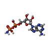  Mass: 426.216 Da / Num. of mol.: 4 / Source method: obtained synthetically / Formula: C10H16N6O9P2 / Feature type: SUBJECT OF INVESTIGATION Mass: 426.216 Da / Num. of mol.: 4 / Source method: obtained synthetically / Formula: C10H16N6O9P2 / Feature type: SUBJECT OF INVESTIGATION#3: Chemical | ChemComp-MG /   Mass: 24.305 Da / Num. of mol.: 4 / Source method: obtained synthetically / Formula: Mg / Feature type: SUBJECT OF INVESTIGATION Mass: 24.305 Da / Num. of mol.: 4 / Source method: obtained synthetically / Formula: Mg / Feature type: SUBJECT OF INVESTIGATION#4: Chemical |   Mass: 92.094 Da / Num. of mol.: 3 / Source method: obtained synthetically / Formula: C3H8O3 / Feature type: SUBJECT OF INVESTIGATION Mass: 92.094 Da / Num. of mol.: 3 / Source method: obtained synthetically / Formula: C3H8O3 / Feature type: SUBJECT OF INVESTIGATION#5: Water | ChemComp-HOH / |  Mass: 18.015 Da / Num. of mol.: 279 / Source method: isolated from a natural source / Formula: H2O Mass: 18.015 Da / Num. of mol.: 279 / Source method: isolated from a natural source / Formula: H2OHas ligand of interest | Y | Has protein modification | Y | |
|---|
-Experimental details
-Experiment
| Experiment | Method: X-RAY DIFFRACTION / Number of used crystals: 1 |
|---|
- Sample preparation
| Crystal | Density Matthews: 2.64 Å3/Da / Density % sol: 53.38 % |
|---|---|
| Crystal grow | Temperature: 293 K / Method: vapor diffusion, sitting drop / pH: 6.5 Details: 0.2 M Magnesium chloride, 0.1 M BIS-TRIS, 25% w/v Polyethylene glycol 3,350 |
-Data collection
| Diffraction | Mean temperature: 100 K / Serial crystal experiment: N |
|---|---|
| Diffraction source | Source: SYNCHROTRON / Site: SSRF / Beamline: BL19U1 / Wavelength: 0.9785 Å |
| Detector | Type: DECTRIS PILATUS3 6M / Detector: PIXEL / Date: Nov 5, 2022 |
| Radiation | Protocol: SINGLE WAVELENGTH / Monochromatic (M) / Laue (L): M / Scattering type: x-ray |
| Radiation wavelength | Wavelength: 0.9785 Å / Relative weight: 1 |
| Reflection | Resolution: 2.3→50 Å / Num. obs: 41783 / % possible obs: 99.7 % / Redundancy: 35.9 % / Biso Wilson estimate: 37.1 Å2 / CC1/2: 0.997 / Rmerge(I) obs: 0.074 / Rrim(I) all: 0.085 / Net I/σ(I): 11 |
| Reflection shell | Resolution: 2.3→2.45 Å / Redundancy: 25.8 % / Rmerge(I) obs: 0.4 / Mean I/σ(I) obs: 2.8 / Num. unique obs: 6076 / CC1/2: 0.903 / Rrim(I) all: 0.479 / % possible all: 99.4 |
- Processing
| Software |
| |||||||||||||||||||||||||||||||||||||||||||||||||||||||||||||||||||||||||||||||||||||||||||||||||||||||||
|---|---|---|---|---|---|---|---|---|---|---|---|---|---|---|---|---|---|---|---|---|---|---|---|---|---|---|---|---|---|---|---|---|---|---|---|---|---|---|---|---|---|---|---|---|---|---|---|---|---|---|---|---|---|---|---|---|---|---|---|---|---|---|---|---|---|---|---|---|---|---|---|---|---|---|---|---|---|---|---|---|---|---|---|---|---|---|---|---|---|---|---|---|---|---|---|---|---|---|---|---|---|---|---|---|---|---|
| Refinement | Method to determine structure: MOLECULAR REPLACEMENT / Resolution: 2.304→34.765 Å / SU ML: 0.3 / Cross valid method: FREE R-VALUE / σ(F): 1.34 / Phase error: 30.69 / Stereochemistry target values: ML
| |||||||||||||||||||||||||||||||||||||||||||||||||||||||||||||||||||||||||||||||||||||||||||||||||||||||||
| Solvent computation | Shrinkage radii: 0.9 Å / VDW probe radii: 1.11 Å / Solvent model: FLAT BULK SOLVENT MODEL | |||||||||||||||||||||||||||||||||||||||||||||||||||||||||||||||||||||||||||||||||||||||||||||||||||||||||
| Refinement step | Cycle: LAST / Resolution: 2.304→34.765 Å
| |||||||||||||||||||||||||||||||||||||||||||||||||||||||||||||||||||||||||||||||||||||||||||||||||||||||||
| Refine LS restraints |
| |||||||||||||||||||||||||||||||||||||||||||||||||||||||||||||||||||||||||||||||||||||||||||||||||||||||||
| LS refinement shell |
|
