 Movie
Movie Controller

[English] 日本語
 Yorodumi
- PDB-8ibs: Crystal structure of GH42 beta-galactosidase BiBga42A from Bifido... -
+ Open data

- Basic information
| Entry | Database: PDB / ID: 8ibs | |||||||||||||||||||||
|---|---|---|---|---|---|---|---|---|---|---|---|---|---|---|---|---|---|---|---|---|---|---|
| Title | Crystal structure of GH42 beta-galactosidase BiBga42A from Bifidobacterium longum subspecies infantis E160A/E318A mutant in complex with galactose | |||||||||||||||||||||
 Components Components | Beta-galactosidase | |||||||||||||||||||||
 Keywords Keywords | HYDROLASE / Glycoside Hydrolase family 42 | |||||||||||||||||||||
| Function / homology |  Function and homology information Function and homology informationbeta-galactosidase complex / beta-galactosidase / beta-galactosidase activity / carbohydrate metabolic process Similarity search - Function | |||||||||||||||||||||
| Biological species |  Bifidobacterium longum subsp. infantis (bacteria) Bifidobacterium longum subsp. infantis (bacteria) | |||||||||||||||||||||
| Method |  X-RAY DIFFRACTION / SYNCHROTRON / MOLECULAR REPLACEMENT / Resolution: 1.9 Å X-RAY DIFFRACTION / SYNCHROTRON / MOLECULAR REPLACEMENT / Resolution: 1.9 Å | |||||||||||||||||||||
 Authors Authors | Hidaka, M. / Fushinobu, S. / Gotoh, A. / Katayama, T. | |||||||||||||||||||||
| Funding support |  Japan, 6items Japan, 6items
| |||||||||||||||||||||
 Citation Citation | Journal: Microbiome Res Rep / Year: 2023 Title: Substrate recognition mode of a glycoside hydrolase family 42 beta-galactosidase from Bifidobacterium longum subspecies infantis ( Bi Bga42A) revealed by crystallographic and mutational analyses. Authors: Gotoh, A. / Hidaka, M. / Sakurama, H. / Nishimoto, M. / Kitaoka, M. / Sakanaka, M. / Fushinobu, S. / Katayama, T. #1: Journal: Glycobiology / Year: 2012 Title: Bifidobacterium longum subsp. infantis uses two different beta-galactosidases for selectively degrading type-1 and type-2 human milk oligosaccharides. Authors: Yoshida, E. / Sakurama, H. / Kiyohara, M. / Nakajima, M. / Kitaoka, M. / Ashida, H. / Hirose, J. / Katayama, T. / Yamamoto, K. / Kumagai, H. | |||||||||||||||||||||
| History |
|
- Structure visualization
| Structure viewer | Molecule: MolmilJmol/JSmol |
|---|
- Downloads & links
-Download
| PDBx/mmCIF format | 8ibs.cif.gz | 848.6 KB | Display | PDBx/mmCIF format |
|---|---|---|---|---|
| PDB format | pdb8ibs.ent.gz | 700.9 KB | Display | PDB format |
| PDBx/mmJSON format | 8ibs.json.gz | Tree view | PDBx/mmJSON format | |
| Others |  Other downloads Other downloads |
-Validation report
| Arichive directory | https://data.pdbj.org/pub/pdb/validation_reports/ib/8ibsftp://data.pdbj.org/pub/pdb/validation_reports/ib/8ibs | HTTPS FTP |
|---|
-Related structure data


-Links
 PDBj
PDBj


- Assembly
| Deposited unit | 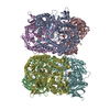
| ||||||||
|---|---|---|---|---|---|---|---|---|---|
| 1 | 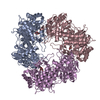
| ||||||||
| 2 | 
| ||||||||
| Unit cell |
|
-Components
| #1: Protein | Mass: 78690.789 Da / Num. of mol.: 6 / Mutation: E160A, E318S Source method: isolated from a genetically manipulated source Source: (gene. exp.) Bifidobacterium longum subsp. infantis (strain ATCC 15697 / DSM 20088 / JCM 1222 / NCTC 11817 / S12) (bacteria)Strain: ATCC 15697 / DSM 20088 / JCM 1222 / NCTC 11817 / S12 Gene: Blon_2016 / Production host: #2: Sugar | ChemComp-GLA / 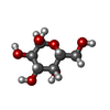  Type: D-saccharide, alpha linking / Mass: 180.156 Da / Num. of mol.: 6 / Source method: obtained synthetically / Formula: C6H12O6 / Feature type: SUBJECT OF INVESTIGATION Type: D-saccharide, alpha linking / Mass: 180.156 Da / Num. of mol.: 6 / Source method: obtained synthetically / Formula: C6H12O6 / Feature type: SUBJECT OF INVESTIGATION#3: Water | ChemComp-HOH / |  Mass: 18.015 Da / Num. of mol.: 2870 / Source method: isolated from a natural source / Formula: H2O Mass: 18.015 Da / Num. of mol.: 2870 / Source method: isolated from a natural source / Formula: H2OHas ligand of interest | Y | |
|---|
-Experimental details
-Experiment
| Experiment | Method: X-RAY DIFFRACTION / Number of used crystals: 1 |
|---|
- Sample preparation
| Crystal | Density Matthews: 2.64 Å3/Da / Density % sol: 53.37 % |
|---|---|
| Crystal grow | Temperature: 293.15 K / Method: vapor diffusion, hanging drop / pH: 6.5 Details: 0.1 M MES-NaOH buffer (pH 6.5), 0.1 M (CH3COO)2Mg, 10% PEG 10000, 25% glycerol, 30 mM lacto-N-tetraose (cleaved) |
-Data collection
| Diffraction | Mean temperature: 100 K / Serial crystal experiment: N |
|---|---|
| Diffraction source | Source: SYNCHROTRON / Site: Photon Factory / Beamline: BL-5A / Wavelength: 1 Å |
| Detector | Type: ADSC QUANTUM 315 / Detector: CCD / Date: Jun 20, 2012 |
| Radiation | Monochromator: Numerical link type Si(111) double crystal / Protocol: SINGLE WAVELENGTH / Monochromatic (M) / Laue (L): M / Scattering type: x-ray |
| Radiation wavelength | Wavelength: 1 Å / Relative weight: 1 |
| Reflection | Resolution: 1.9→48.57 Å / Num. obs: 376638 / % possible obs: 98.3 % / Redundancy: 3.7 % / Biso Wilson estimate: 22.06 Å2 / CC1/2: 0.997 / Rmerge(I) obs: 0.097 / Rpim(I) all: 0.058 / Net I/σ(I): 9.5 |
| Reflection shell | Resolution: 1.9→1.93 Å / Redundancy: 2.8 % / Rmerge(I) obs: 0.935 / Mean I/σ(I) obs: 1.1 / Num. unique obs: 15738 / CC1/2: 0.545 / Rpim(I) all: 0.672 / % possible all: 83 |
- Processing
| Software |
| ||||||||||||||||||||||||||||||||||||||||||||||||||||||||||||||||||||||||||||||||||||||||||||||||||||||||||||||||||||||||||||||||||||||||||||||||||||||||||||||||||||||||||||||||||||||
|---|---|---|---|---|---|---|---|---|---|---|---|---|---|---|---|---|---|---|---|---|---|---|---|---|---|---|---|---|---|---|---|---|---|---|---|---|---|---|---|---|---|---|---|---|---|---|---|---|---|---|---|---|---|---|---|---|---|---|---|---|---|---|---|---|---|---|---|---|---|---|---|---|---|---|---|---|---|---|---|---|---|---|---|---|---|---|---|---|---|---|---|---|---|---|---|---|---|---|---|---|---|---|---|---|---|---|---|---|---|---|---|---|---|---|---|---|---|---|---|---|---|---|---|---|---|---|---|---|---|---|---|---|---|---|---|---|---|---|---|---|---|---|---|---|---|---|---|---|---|---|---|---|---|---|---|---|---|---|---|---|---|---|---|---|---|---|---|---|---|---|---|---|---|---|---|---|---|---|---|---|---|---|---|
| Refinement | Method to determine structure: MOLECULAR REPLACEMENT / Resolution: 1.9→48.61 Å / Cor.coef. Fo:Fc: 0.959 / Cor.coef. Fo:Fc free: 0.937 / SU B: 4.266 / SU ML: 0.118 / Cross valid method: THROUGHOUT / ESU R: 0.148 / ESU R Free: 0.14 / Stereochemistry target values: MAXIMUM LIKELIHOOD / Details: HYDROGENS HAVE BEEN ADDED IN THE RIDING POSITIONS
| ||||||||||||||||||||||||||||||||||||||||||||||||||||||||||||||||||||||||||||||||||||||||||||||||||||||||||||||||||||||||||||||||||||||||||||||||||||||||||||||||||||||||||||||||||||||
| Solvent computation | Ion probe radii: 0.8 Å / Shrinkage radii: 0.8 Å / VDW probe radii: 1.2 Å / Solvent model: MASK | ||||||||||||||||||||||||||||||||||||||||||||||||||||||||||||||||||||||||||||||||||||||||||||||||||||||||||||||||||||||||||||||||||||||||||||||||||||||||||||||||||||||||||||||||||||||
| Displacement parameters | Biso mean: 24.001 Å2
| ||||||||||||||||||||||||||||||||||||||||||||||||||||||||||||||||||||||||||||||||||||||||||||||||||||||||||||||||||||||||||||||||||||||||||||||||||||||||||||||||||||||||||||||||||||||
| Refinement step | Cycle: 1 / Resolution: 1.9→48.61 Å
| ||||||||||||||||||||||||||||||||||||||||||||||||||||||||||||||||||||||||||||||||||||||||||||||||||||||||||||||||||||||||||||||||||||||||||||||||||||||||||||||||||||||||||||||||||||||
| Refine LS restraints |
|