 Movie
Movie Controller

[English] 日本語
 Yorodumi
- PDB-8ibl: MES bound form of PET-degrading cutinase Cut190 with thermostabil... -
+ Open data

- Basic information
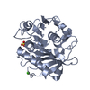
| Entry | Database: PDB / ID: 8ibl | ||||||
|---|---|---|---|---|---|---|---|
| Title | MES bound form of PET-degrading cutinase Cut190 with thermostability-improving mutations of S226P/R228S/Q138A/D250C-E296C/Q123H/N202H and S176A inactivation | ||||||
 Components Components | Alpha/beta hydrolase family protein | ||||||
 Keywords Keywords | HYDROLASE / PROTEIN ENGINEERING / POLYESTERASE / DISULFIDE BOND / METAL BINDING / Ligand complex | ||||||
| Function / homology | Platelet-activating factor acetylhydrolase, isoform II / cutinase / carboxylic ester hydrolase activity / Alpha/Beta hydrolase fold / metal ion binding / cutinase Function and homology information Function and homology information | ||||||
| Biological species |  Saccharomonospora viridis (bacteria) Saccharomonospora viridis (bacteria) | ||||||
| Method |  X-RAY DIFFRACTION / SYNCHROTRON / MOLECULAR REPLACEMENT / Resolution: 2.6 Å X-RAY DIFFRACTION / SYNCHROTRON / MOLECULAR REPLACEMENT / Resolution: 2.6 Å | ||||||
 Authors Authors | Emori, M. / Numoto, N. / Kamiya, N. / Oda, M. | ||||||
| Funding support |  Japan, 1items Japan, 1items
| ||||||
 Citation Citation | Journal: Biorxiv / Year: 2023 Title: Improvement of thermostability and activity of PET-degrading enzyme Cut190 towards a detailed understanding and application of the enzymatic reaction mechanism. Authors: Numoto, N. / Kamiya, N. / Oda, M. | ||||||
| History |
|
- Structure visualization
| Structure viewer | Molecule: MolmilJmol/JSmol |
|---|
- Downloads & links
-Download
| PDBx/mmCIF format | 8ibl.cif.gz | 120.8 KB | Display | PDBx/mmCIF format |
|---|---|---|---|---|
| PDB format | pdb8ibl.ent.gz | 87.5 KB | Display | PDB format |
| PDBx/mmJSON format | 8ibl.json.gz | Tree view | PDBx/mmJSON format | |
| Others |  Other downloads Other downloads |
-Validation report
| Summary document | 8ibl_validation.pdf.gz | 970.2 KB | Display | wwPDB validaton report |
|---|---|---|---|---|
| Full document | 8ibl_full_validation.pdf.gz | 963.8 KB | Display | |
| Data in XML | 8ibl_validation.xml.gz | 20.5 KB | Display | |
| Data in CIF | 8ibl_validation.cif.gz | 27.9 KB | Display | |
| Arichive directory | https://data.pdbj.org/pub/pdb/validation_reports/ib/8iblftp://data.pdbj.org/pub/pdb/validation_reports/ib/8ibl | HTTPS FTP |
-Related structure data
-Links
 PDBj
PDBj
- Assembly
| Deposited unit | 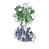
| ||||||||||||
|---|---|---|---|---|---|---|---|---|---|---|---|---|---|
| 1 | 
| ||||||||||||
| 2 | 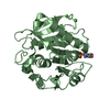
| ||||||||||||
| Unit cell |
|
-Components
| #1: Protein | Mass: 28889.537 Da / Num. of mol.: 2 Mutation: Q123H,Q138A, S176A, N202H, S226P, R228S, D250C, E296C Source method: isolated from a genetically manipulated source Source: (gene. exp.) Saccharomonospora viridis (bacteria) / Gene: Cut190, SAMN02982918_2340 / Production host: #2: Chemical |   Mass: 40.078 Da / Num. of mol.: 2 / Source method: obtained synthetically / Formula: Ca Mass: 40.078 Da / Num. of mol.: 2 / Source method: obtained synthetically / Formula: Ca#3: Chemical |   Mass: 195.237 Da / Num. of mol.: 2 / Source method: obtained synthetically / Formula: C6H13NO4S / Feature type: SUBJECT OF INVESTIGATION / Comment: pH buffer*YM Mass: 195.237 Da / Num. of mol.: 2 / Source method: obtained synthetically / Formula: C6H13NO4S / Feature type: SUBJECT OF INVESTIGATION / Comment: pH buffer*YM#4: Chemical |   Mass: 35.453 Da / Num. of mol.: 2 / Source method: obtained synthetically / Formula: Cl Mass: 35.453 Da / Num. of mol.: 2 / Source method: obtained synthetically / Formula: Cl#5: Water | ChemComp-HOH / |  Mass: 18.015 Da / Num. of mol.: 61 / Source method: isolated from a natural source / Formula: H2O Mass: 18.015 Da / Num. of mol.: 61 / Source method: isolated from a natural source / Formula: H2OHas ligand of interest | Y | |
|---|
-Experimental details
-Experiment
| Experiment | Method: X-RAY DIFFRACTION / Number of used crystals: 1 |
|---|
- Sample preparation
| Crystal | Density Matthews: 2.27 Å3/Da / Density % sol: 45.92 % |
|---|---|
| Crystal grow | Temperature: 293 K / Method: vapor diffusion, hanging drop / pH: 6.5 Details: 0.1 M MES monohydrate pH 6.5, 0.2 M Ammonium sulfate, and 30% (w/v) PEG MME 5,000 |
-Data collection
| Diffraction | Mean temperature: 95 K / Serial crystal experiment: N |
|---|---|
| Diffraction source | Source: SYNCHROTRON / Site: Photon Factory / Beamline: BL-17A / Wavelength: 0.9 Å |
| Detector | Type: DECTRIS EIGER X 16M / Detector: PIXEL / Date: Mar 9, 2021 |
| Radiation | Protocol: SINGLE WAVELENGTH / Monochromatic (M) / Laue (L): M / Scattering type: x-ray |
| Radiation wavelength | Wavelength: 0.9 Å / Relative weight: 1 |
| Reflection | Resolution: 2.6→33 Å / Num. obs: 15681 / % possible obs: 99.9 % / Redundancy: 5.4 % / Biso Wilson estimate: 36.34 Å2 / CC1/2: 0.98 / Rsym value: 0.276 / Net I/σ(I): 5.8 |
| Reflection shell | Resolution: 2.61→2.76 Å / Num. unique obs: 2506 / CC1/2: 0.642 / Rsym value: 1.22 |
- Processing
| Software |
| |||||||||||||||||||||||||||||||||||||||||||||||||
|---|---|---|---|---|---|---|---|---|---|---|---|---|---|---|---|---|---|---|---|---|---|---|---|---|---|---|---|---|---|---|---|---|---|---|---|---|---|---|---|---|---|---|---|---|---|---|---|---|---|---|
| Refinement | Method to determine structure: MOLECULAR REPLACEMENT / Resolution: 2.6→32.36 Å / SU ML: 0.3911 / Cross valid method: FREE R-VALUE / σ(F): 1.98 / Phase error: 29.2956 Stereochemistry target values: GeoStd + Monomer Library + CDL v1.2
| |||||||||||||||||||||||||||||||||||||||||||||||||
| Solvent computation | Shrinkage radii: 0.9 Å / VDW probe radii: 1.11 Å / Solvent model: FLAT BULK SOLVENT MODEL | |||||||||||||||||||||||||||||||||||||||||||||||||
| Displacement parameters | Biso mean: 36.35 Å2 | |||||||||||||||||||||||||||||||||||||||||||||||||
| Refinement step | Cycle: LAST / Resolution: 2.6→32.36 Å
| |||||||||||||||||||||||||||||||||||||||||||||||||
| Refine LS restraints |
| |||||||||||||||||||||||||||||||||||||||||||||||||
| LS refinement shell |
|
