 Movie
Movie Controller

[English] 日本語
 Yorodumi
- PDB-8ia1: plastidial glycerol-3-phosphate acyltransferases (GPAT) from the ... -
+ Open data

- Basic information
| Entry | Database: PDB / ID: 8ia1 | ||||||
|---|---|---|---|---|---|---|---|
| Title | plastidial glycerol-3-phosphate acyltransferases (GPAT) from the green alga Myrmecia incisa | ||||||
 Components Components | Glycerol-3-phosphate acyltransferase, chloroplastic | ||||||
 Keywords Keywords | TRANSFERASE / plastidial glycerol-3-phosphate acyltransferases | ||||||
| Function / homology |  Function and homology information Function and homology information: / glycerol-3-phosphate 1-O-acyltransferase / glycerol-3-phosphate O-acyltransferase activity / CDP-diacylglycerol biosynthetic process / chloroplast stroma Similarity search - Function | ||||||
| Biological species |  Lobosphaera incisa (plant) Lobosphaera incisa (plant) | ||||||
| Method |  X-RAY DIFFRACTION / SYNCHROTRON / MOLECULAR REPLACEMENT / Resolution: 2.31 Å X-RAY DIFFRACTION / SYNCHROTRON / MOLECULAR REPLACEMENT / Resolution: 2.31 Å | ||||||
 Authors Authors | Song, X. | ||||||
| Funding support |  China, 1items China, 1items
| ||||||
 Citation Citation | Journal: Plant Sci. / Year: 2023 Title: Unique recognition of the microalgal plastidial glycerol-3-phosphate acyltransferase for acyl-ACP. Authors: Li, X. / Yang, M. / Sun, D. / Shi, J. / Yang, M. / Feng, Y. / Xue, S. | ||||||
| History |
|
- Structure visualization
| Structure viewer | Molecule: MolmilJmol/JSmol |
|---|
- Downloads & links
-Download
| PDBx/mmCIF format | 8ia1.cif.gz | 275.8 KB | Display | PDBx/mmCIF format |
|---|---|---|---|---|
| PDB format | pdb8ia1.ent.gz | 180.7 KB | Display | PDB format |
| PDBx/mmJSON format | 8ia1.json.gz | Tree view | PDBx/mmJSON format | |
| Others |  Other downloads Other downloads |
-Validation report
| Summary document | 8ia1_validation.pdf.gz | 444.1 KB | Display | wwPDB validaton report |
|---|---|---|---|---|
| Full document | 8ia1_full_validation.pdf.gz | 453.6 KB | Display | |
| Data in XML | 8ia1_validation.xml.gz | 40.8 KB | Display | |
| Data in CIF | 8ia1_validation.cif.gz | 58.3 KB | Display | |
| Arichive directory | https://data.pdbj.org/pub/pdb/validation_reports/ia/8ia1ftp://data.pdbj.org/pub/pdb/validation_reports/ia/8ia1 | HTTPS FTP |
-Related structure data
| Similar structure data |
|---|
-Links
 PDBj
PDBj


- Assembly
| Deposited unit | 
| ||||||||||||
|---|---|---|---|---|---|---|---|---|---|---|---|---|---|
| 1 | 
| ||||||||||||
| 2 | 
| ||||||||||||
| 3 | 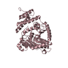
| ||||||||||||
| Unit cell |
|
-Components
| #1: Protein | Mass: 41651.180 Da / Num. of mol.: 3 Source method: isolated from a genetically manipulated source Source: (gene. exp.) Lobosphaera incisa (plant) / Gene: GPAT / Production host:  #2: Water | ChemComp-HOH / |  Mass: 18.015 Da / Num. of mol.: 351 / Source method: isolated from a natural source / Formula: H2O Mass: 18.015 Da / Num. of mol.: 351 / Source method: isolated from a natural source / Formula: H2OHas protein modification | N | |
|---|
-Experimental details
-Experiment
| Experiment | Method: X-RAY DIFFRACTION / Number of used crystals: 1 |
|---|
- Sample preparation
| Crystal | Density Matthews: 2.51 Å3/Da / Density % sol: 51.04 % |
|---|---|
| Crystal grow | Temperature: 289 K / Method: vapor diffusion, hanging drop Details: 0.2M (NH4)2SO4, 0.1M Tris-HCl pH 8.5, 20% (w/v) PEG 3350 |
-Data collection
| Diffraction | Mean temperature: 100 K / Serial crystal experiment: N |
|---|---|
| Diffraction source | Source: SYNCHROTRON / Site: SSRF / Beamline: BL18U1 / Wavelength: 1.0001 Å |
| Detector | Type: MAR CCD 165 mm / Detector: CCD / Date: Dec 7, 2017 |
| Radiation | Protocol: SINGLE WAVELENGTH / Monochromatic (M) / Laue (L): M / Scattering type: x-ray |
| Radiation wavelength | Wavelength: 1.0001 Å / Relative weight: 1 |
| Reflection | Resolution: 2.31→37.32 Å / Num. obs: 50682 / % possible obs: 89.19 % / Redundancy: 2.2 % / Biso Wilson estimate: 28.13 Å2 / CC1/2: 0.99 / Net I/av σ(I): 16.1 / Net I/σ(I): 16.1 |
| Reflection shell | Resolution: 2.31→2.38 Å / Mean I/σ(I) obs: 2.61 / Num. unique obs: 2902 / CC1/2: 0.76 |
- Processing
| Software |
| ||||||||||||||||||||||||||||||||||||||||||||||||||||||||||||||||||||||||||||||||||||||||||||||||||||||||||||||||||||||||||||||
|---|---|---|---|---|---|---|---|---|---|---|---|---|---|---|---|---|---|---|---|---|---|---|---|---|---|---|---|---|---|---|---|---|---|---|---|---|---|---|---|---|---|---|---|---|---|---|---|---|---|---|---|---|---|---|---|---|---|---|---|---|---|---|---|---|---|---|---|---|---|---|---|---|---|---|---|---|---|---|---|---|---|---|---|---|---|---|---|---|---|---|---|---|---|---|---|---|---|---|---|---|---|---|---|---|---|---|---|---|---|---|---|---|---|---|---|---|---|---|---|---|---|---|---|---|---|---|---|
| Refinement | Method to determine structure: MOLECULAR REPLACEMENT / Resolution: 2.31→37.32 Å / SU ML: 0.2843 / Cross valid method: FREE R-VALUE / σ(F): 1.99 / Phase error: 24.9081 Stereochemistry target values: GeoStd + Monomer Library + CDL v1.2
| ||||||||||||||||||||||||||||||||||||||||||||||||||||||||||||||||||||||||||||||||||||||||||||||||||||||||||||||||||||||||||||||
| Solvent computation | Shrinkage radii: 0.9 Å / VDW probe radii: 1.11 Å / Solvent model: FLAT BULK SOLVENT MODEL | ||||||||||||||||||||||||||||||||||||||||||||||||||||||||||||||||||||||||||||||||||||||||||||||||||||||||||||||||||||||||||||||
| Displacement parameters | Biso mean: 33.65 Å2 | ||||||||||||||||||||||||||||||||||||||||||||||||||||||||||||||||||||||||||||||||||||||||||||||||||||||||||||||||||||||||||||||
| Refinement step | Cycle: LAST / Resolution: 2.31→37.32 Å
| ||||||||||||||||||||||||||||||||||||||||||||||||||||||||||||||||||||||||||||||||||||||||||||||||||||||||||||||||||||||||||||||
| Refine LS restraints |
| ||||||||||||||||||||||||||||||||||||||||||||||||||||||||||||||||||||||||||||||||||||||||||||||||||||||||||||||||||||||||||||||
| LS refinement shell |
|
