 Movie
Movie Controller

[English] 日本語
 Yorodumi
- PDB-8i5j: Crystal structure of chitin oligosaccharide binding protein from ... -
+ Open data

- Basic information
| Entry | Database: PDB / ID: 8i5j | ||||||
|---|---|---|---|---|---|---|---|
| Title | Crystal structure of chitin oligosaccharide binding protein from Vibrio cholera. | ||||||
 Components Components | ABC transporter substrate-binding protein | ||||||
 Keywords Keywords | SUGAR BINDING PROTEIN / chitin oligosaccharide / periplasmic / solute binding protein | ||||||
| Function / homology |  Function and homology information Function and homology informationdipeptide transport / peptide transmembrane transporter activity / ATP-binding cassette (ABC) transporter complex / outer membrane-bounded periplasmic space / metal ion binding Similarity search - Function | ||||||
| Biological species |   Vibrio cholerae (bacteria) Vibrio cholerae (bacteria) | ||||||
| Method |  X-RAY DIFFRACTION / SYNCHROTRON / MOLECULAR REPLACEMENT / Resolution: 1.602 Å X-RAY DIFFRACTION / SYNCHROTRON / MOLECULAR REPLACEMENT / Resolution: 1.602 Å | ||||||
 Authors Authors | Ohnuma, T. / Takeshita, D. | ||||||
| Funding support | 1items
| ||||||
 Citation Citation | Journal: Sci Rep / Year: 2023 Title: Periplasmic chitooligosaccharide-binding protein requires a three-domain organization for substrate translocation. Authors: Ohnuma, T. / Tsujii, J. / Kataoka, C. / Yoshimoto, T. / Takeshita, D. / Lampela, O. / Juffer, A.H. / Suginta, W. / Fukamizo, T. | ||||||
| History |
|
- Structure visualization
| Structure viewer | Molecule: MolmilJmol/JSmol |
|---|
- Downloads & links
-Download
| PDBx/mmCIF format | 8i5j.cif.gz | 237.9 KB | Display | PDBx/mmCIF format |
|---|---|---|---|---|
| PDB format | pdb8i5j.ent.gz | 185.8 KB | Display | PDB format |
| PDBx/mmJSON format | 8i5j.json.gz | Tree view | PDBx/mmJSON format | |
| Others |  Other downloads Other downloads |
-Validation report
| Summary document | 8i5j_validation.pdf.gz | 431.8 KB | Display | wwPDB validaton report |
|---|---|---|---|---|
| Full document | 8i5j_full_validation.pdf.gz | 433.9 KB | Display | |
| Data in XML | 8i5j_validation.xml.gz | 43.4 KB | Display | |
| Data in CIF | 8i5j_validation.cif.gz | 65.3 KB | Display | |
| Arichive directory | https://data.pdbj.org/pub/pdb/validation_reports/i5/8i5jftp://data.pdbj.org/pub/pdb/validation_reports/i5/8i5j | HTTPS FTP |
-Related structure data
| Related structure data | 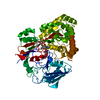 8i5kC 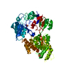 4gf8S S: Starting model for refinement C: citing same article ( |
|---|---|
| Similar structure data |
-Links
 PDBj
PDBj- Assembly
| Deposited unit | 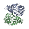
| ||||||||
|---|---|---|---|---|---|---|---|---|---|
| 1 | 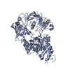
| ||||||||
| 2 | 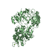
| ||||||||
| Unit cell |
|
-Components
| #1: Protein | Mass: 60977.188 Da / Num. of mol.: 2 Source method: isolated from a genetically manipulated source Source: (gene. exp.) Vibrio cholerae (bacteria)Gene: appA_1, appA, D6U24_16190, ERS013201_01979, ERS013202_02445, EYB64_10090, F0H40_11710, F0M16_04225, FLM12_17525 Production host: #2: Chemical |   Mass: 24.305 Da / Num. of mol.: 2 / Source method: obtained synthetically / Formula: Mg Mass: 24.305 Da / Num. of mol.: 2 / Source method: obtained synthetically / Formula: Mg#3: Water | ChemComp-HOH / |  Mass: 18.015 Da / Num. of mol.: 773 / Source method: isolated from a natural source / Formula: H2O Mass: 18.015 Da / Num. of mol.: 773 / Source method: isolated from a natural source / Formula: H2OHas ligand of interest | N | Has protein modification | Y | |
|---|
-Experimental details
-Experiment
| Experiment | Method: X-RAY DIFFRACTION / Number of used crystals: 1 |
|---|
- Sample preparation
| Crystal | Density Matthews: 2.3 Å3/Da / Density % sol: 46.47 % |
|---|---|
| Crystal grow | Temperature: 293 K / Method: vapor diffusion, sitting drop / pH: 5.6 Details: 0.2 M Ammonium acetate, 0.1 M Sodium citrate tribasic dehydrate, Polyethylene glycol 4,000 |
-Data collection
| Diffraction | Mean temperature: 100 K / Serial crystal experiment: N |
|---|---|
| Diffraction source | Source: SYNCHROTRON / Site: Photon Factory  / Beamline: BL-17A / Wavelength: 0.98 Å / Beamline: BL-17A / Wavelength: 0.98 Å |
| Detector | Type: DECTRIS EIGER X 16M / Detector: PIXEL / Date: Dec 19, 2017 |
| Radiation | Protocol: SINGLE WAVELENGTH / Monochromatic (M) / Laue (L): M / Scattering type: x-ray |
| Radiation wavelength | Wavelength: 0.98 Å / Relative weight: 1 |
| Reflection | Resolution: 1.602→19.81 Å / Num. obs: 261371 / % possible obs: 91.63 % / Redundancy: 3.27 % / CC1/2: 0.994 / Net I/σ(I): 8.75 |
| Reflection shell | Resolution: 1.602→1.69 Å / Num. unique obs: 28401 / CC1/2: 0.519 |
- Processing
| Software |
| |||||||||||||||||||||||||||||||||||||||||||||||||||||||||||||||||||||||||||||||||||||||||||||||||||||||||||||||||||||||||||||||||||||||||||||||||||||||||||||||||||||||||||||||||||||||||||||||||||||||||||||||||||||||||
|---|---|---|---|---|---|---|---|---|---|---|---|---|---|---|---|---|---|---|---|---|---|---|---|---|---|---|---|---|---|---|---|---|---|---|---|---|---|---|---|---|---|---|---|---|---|---|---|---|---|---|---|---|---|---|---|---|---|---|---|---|---|---|---|---|---|---|---|---|---|---|---|---|---|---|---|---|---|---|---|---|---|---|---|---|---|---|---|---|---|---|---|---|---|---|---|---|---|---|---|---|---|---|---|---|---|---|---|---|---|---|---|---|---|---|---|---|---|---|---|---|---|---|---|---|---|---|---|---|---|---|---|---|---|---|---|---|---|---|---|---|---|---|---|---|---|---|---|---|---|---|---|---|---|---|---|---|---|---|---|---|---|---|---|---|---|---|---|---|---|---|---|---|---|---|---|---|---|---|---|---|---|---|---|---|---|---|---|---|---|---|---|---|---|---|---|---|---|---|---|---|---|---|---|---|---|---|---|---|---|---|---|---|---|---|---|---|---|---|
| Refinement | Method to determine structure: MOLECULAR REPLACEMENT Starting model: 4GF8 Resolution: 1.602→19.81 Å / SU ML: 0.16 / Cross valid method: FREE R-VALUE / σ(F): 1.02 / Phase error: 20.42 / Stereochemistry target values: ML
| |||||||||||||||||||||||||||||||||||||||||||||||||||||||||||||||||||||||||||||||||||||||||||||||||||||||||||||||||||||||||||||||||||||||||||||||||||||||||||||||||||||||||||||||||||||||||||||||||||||||||||||||||||||||||
| Solvent computation | Shrinkage radii: 0.9 Å / VDW probe radii: 1.11 Å / Solvent model: FLAT BULK SOLVENT MODEL | |||||||||||||||||||||||||||||||||||||||||||||||||||||||||||||||||||||||||||||||||||||||||||||||||||||||||||||||||||||||||||||||||||||||||||||||||||||||||||||||||||||||||||||||||||||||||||||||||||||||||||||||||||||||||
| Refinement step | Cycle: LAST / Resolution: 1.602→19.81 Å
| |||||||||||||||||||||||||||||||||||||||||||||||||||||||||||||||||||||||||||||||||||||||||||||||||||||||||||||||||||||||||||||||||||||||||||||||||||||||||||||||||||||||||||||||||||||||||||||||||||||||||||||||||||||||||
| Refine LS restraints |
| |||||||||||||||||||||||||||||||||||||||||||||||||||||||||||||||||||||||||||||||||||||||||||||||||||||||||||||||||||||||||||||||||||||||||||||||||||||||||||||||||||||||||||||||||||||||||||||||||||||||||||||||||||||||||
| LS refinement shell |
|
