 Movie
Movie Controller

[English] 日本語
 Yorodumi
- PDB-8i3p: crystal structure of yeast cytosine deaminase mutant yCD-RQ-1/8SA... -
+ Open data

- Basic information
| Entry | Database: PDB / ID: 8i3p | ||||||
|---|---|---|---|---|---|---|---|
| Title | crystal structure of yeast cytosine deaminase mutant yCD-RQ-1/8SAH in complex with (R)-4-hydroxy-3,4-dihydropyrimidin-2(1H)-one | ||||||
 Components Components | Cytosine deaminase | ||||||
 Keywords Keywords | HYDROLASE / metalloenzyme | ||||||
| Function / homology |  Function and homology information Function and homology informationcytidine metabolic process / pyrimidine-containing compound salvage / diaminohydroxyphosphoribosylaminopyrimidine deaminase activity / cytosine deaminase / cytosine deaminase activity / UMP salvage / cytosine metabolic process / zinc ion binding / nucleus / cytoplasm Similarity search - Function | ||||||
| Biological species |  | ||||||
| Method |  X-RAY DIFFRACTION / SYNCHROTRON / MOLECULAR REPLACEMENT / Resolution: 1.3 Å X-RAY DIFFRACTION / SYNCHROTRON / MOLECULAR REPLACEMENT / Resolution: 1.3 Å | ||||||
 Authors Authors | Qin, M.M. / Deng, H.Z. / Yao, L.S. | ||||||
| Funding support |  China, 1items China, 1items
| ||||||
 Citation Citation | Journal: To be published Title: crystal structure of yeast cytosine deaminase mutant yCD-RQ-1/8SAH in complex with (R)-4-hydroxy-3,4-dihydropyrimidin-2(1H)-one Authors: Qin, M.M. / Deng, H.Z. / Yao, L.S. | ||||||
| History |
|
- Structure visualization
| Structure viewer | Molecule: MolmilJmol/JSmol |
|---|
- Downloads & links
-Download
| PDBx/mmCIF format | 8i3p.cif.gz | 87 KB | Display | PDBx/mmCIF format |
|---|---|---|---|---|
| PDB format | pdb8i3p.ent.gz | 62.9 KB | Display | PDB format |
| PDBx/mmJSON format | 8i3p.json.gz | Tree view | PDBx/mmJSON format | |
| Others |  Other downloads Other downloads |
-Validation report
| Arichive directory | https://data.pdbj.org/pub/pdb/validation_reports/i3/8i3pftp://data.pdbj.org/pub/pdb/validation_reports/i3/8i3p | HTTPS FTP |
|---|
-Related structure data
| Similar structure data |
|---|
-Links
 PDBj
PDBj- Assembly
| Deposited unit | 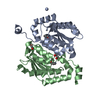
| ||||||||
|---|---|---|---|---|---|---|---|---|---|
| 1 |
| ||||||||
| Unit cell |
|
-Components
| #1: Protein | Mass: 22764.852 Da / Num. of mol.: 2 Source method: isolated from a genetically manipulated source Source: (gene. exp.)  #2: Chemical |   Mass: 114.103 Da / Num. of mol.: 2 / Source method: obtained synthetically / Formula: C4H6N2O2 / Feature type: SUBJECT OF INVESTIGATION Mass: 114.103 Da / Num. of mol.: 2 / Source method: obtained synthetically / Formula: C4H6N2O2 / Feature type: SUBJECT OF INVESTIGATION#3: Chemical |   Mass: 65.409 Da / Num. of mol.: 2 / Source method: obtained synthetically / Formula: Zn Mass: 65.409 Da / Num. of mol.: 2 / Source method: obtained synthetically / Formula: Zn#4: Chemical |   Mass: 62.068 Da / Num. of mol.: 2 / Source method: isolated from a natural source / Formula: C2H6O2 Mass: 62.068 Da / Num. of mol.: 2 / Source method: isolated from a natural source / Formula: C2H6O2#5: Water | ChemComp-HOH / |  Mass: 18.015 Da / Num. of mol.: 349 / Source method: isolated from a natural source / Formula: H2O Mass: 18.015 Da / Num. of mol.: 349 / Source method: isolated from a natural source / Formula: H2OHas ligand of interest | Y | |
|---|
-Experimental details
-Experiment
| Experiment | Method: X-RAY DIFFRACTION / Number of used crystals: 1 |
|---|
- Sample preparation
| Crystal | Density Matthews: 1.96 Å3/Da / Density % sol: 31.73 % |
|---|---|
| Crystal grow | Temperature: 291 K / Method: vapor diffusion, sitting drop Details: 1.5 M Ammonium sulfate, 0.1 M Tris pH 8.5, 12% v/v Glycerol |
-Data collection
| Diffraction | Mean temperature: 100 K / Serial crystal experiment: N |
|---|---|
| Diffraction source | Source: SYNCHROTRON / Site: SSRF / Beamline: BL10U2 / Wavelength: 0.979 Å |
| Detector | Type: DECTRIS EIGER X 16M / Detector: PIXEL / Date: Aug 21, 2022 |
| Radiation | Protocol: SINGLE WAVELENGTH / Monochromatic (M) / Laue (L): M / Scattering type: x-ray |
| Radiation wavelength | Wavelength: 0.979 Å / Relative weight: 1 |
| Reflection | Resolution: 1.18→48.42 Å / Num. obs: 69899 / % possible obs: 85.6 % / Redundancy: 2.5 % / CC1/2: 0.998 / Rmerge(I) obs: 0.042 / Rpim(I) all: 0.031 / Rrim(I) all: 0.053 / Χ2: 0.46 / Net I/σ(I): 9.5 / Num. measured all: 223856 |
| Reflection shell | Resolution: 1.18→1.24 Å / % possible obs: 81.1 % / Redundancy: 2.2 % / Rmerge(I) obs: 0.262 / Num. measured all: 27029 / Num. unique obs: 12501 / CC1/2: 0.902 / Rpim(I) all: 0.22 / Rrim(I) all: 0.344 / Χ2: 0.39 / Net I/σ(I) obs: 2.5 |
- Processing
| Software |
| ||||||||||||||||||||||||||||||||||||||||||||||||||||||||||||||||||||||||||||||||||||
|---|---|---|---|---|---|---|---|---|---|---|---|---|---|---|---|---|---|---|---|---|---|---|---|---|---|---|---|---|---|---|---|---|---|---|---|---|---|---|---|---|---|---|---|---|---|---|---|---|---|---|---|---|---|---|---|---|---|---|---|---|---|---|---|---|---|---|---|---|---|---|---|---|---|---|---|---|---|---|---|---|---|---|---|---|---|
| Refinement | Method to determine structure: MOLECULAR REPLACEMENT / Resolution: 1.3→33.41 Å / SU ML: 0.09 / Cross valid method: THROUGHOUT / σ(F): 1.34 / Phase error: 15.77 / Stereochemistry target values: ML
| ||||||||||||||||||||||||||||||||||||||||||||||||||||||||||||||||||||||||||||||||||||
| Solvent computation | Shrinkage radii: 0.9 Å / VDW probe radii: 1.11 Å / Solvent model: FLAT BULK SOLVENT MODEL | ||||||||||||||||||||||||||||||||||||||||||||||||||||||||||||||||||||||||||||||||||||
| Refinement step | Cycle: LAST / Resolution: 1.3→33.41 Å
| ||||||||||||||||||||||||||||||||||||||||||||||||||||||||||||||||||||||||||||||||||||
| Refine LS restraints |
| ||||||||||||||||||||||||||||||||||||||||||||||||||||||||||||||||||||||||||||||||||||
| LS refinement shell |
|
