 Movie
Movie Controller

[English] 日本語
 Yorodumi
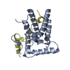
- PDB-8i3f: Crystal structure of Rco1-Eaf3 with peptide of histone H3 N-terminal -
+ Open data

- Basic information
| Entry | Database: PDB / ID: 8i3f | ||||||
|---|---|---|---|---|---|---|---|
| Title | Crystal structure of Rco1-Eaf3 with peptide of histone H3 N-terminal | ||||||
 Components Components |
| ||||||
 Keywords Keywords | STRUCTURAL PROTEIN / MRG domain / complex / PHD domain | ||||||
| Function / homology |  Function and homology information Function and homology informationRpd3S complex / NuA4 histone acetyltransferase complex / chromatin remodeling / DNA repair / regulation of DNA-templated transcription / regulation of transcription by RNA polymerase II / metal ion binding Similarity search - Function | ||||||
| Biological species |  | ||||||
| Method |  X-RAY DIFFRACTION / SYNCHROTRON / MOLECULAR REPLACEMENT / Resolution: 1.62 Å X-RAY DIFFRACTION / SYNCHROTRON / MOLECULAR REPLACEMENT / Resolution: 1.62 Å | ||||||
 Authors Authors | Chen, Z. / Xu, C. | ||||||
| Funding support |  China, 1items China, 1items
| ||||||
 Citation Citation | Journal: Cell Discov / Year: 2023 Title: Molecular basis for Eaf3-mediated assembly of Rpd3S and NuA4. Authors: Chen, Z. / Lundy, T. / Zhu, Z. / Hoskins, V.E. / Zhang, J. / Yao, X. / Strahl, B.D. / Xu, C. | ||||||
| History |
|
- Structure visualization
| Structure viewer | Molecule: MolmilJmol/JSmol |
|---|
- Downloads & links
-Download
| PDBx/mmCIF format | 8i3f.cif.gz | 143.7 KB | Display | PDBx/mmCIF format |
|---|---|---|---|---|
| PDB format | pdb8i3f.ent.gz | 110.7 KB | Display | PDB format |
| PDBx/mmJSON format | 8i3f.json.gz | Tree view | PDBx/mmJSON format | |
| Others |  Other downloads Other downloads |
-Validation report
| Summary document | 8i3f_validation.pdf.gz | 1.7 MB | Display | wwPDB validaton report |
|---|---|---|---|---|
| Full document | 8i3f_full_validation.pdf.gz | 1.7 MB | Display | |
| Data in XML | 8i3f_validation.xml.gz | 16.1 KB | Display | |
| Data in CIF | 8i3f_validation.cif.gz | 23.6 KB | Display | |
| Arichive directory | https://data.pdbj.org/pub/pdb/validation_reports/i3/8i3fftp://data.pdbj.org/pub/pdb/validation_reports/i3/8i3f | HTTPS FTP |
-Related structure data
-Links
 PDBj
PDBj- Assembly
| Deposited unit | 
| ||||||||
|---|---|---|---|---|---|---|---|---|---|
| 1 |
| ||||||||
| Unit cell |
|
-Components
| #1: Protein | Mass: 21127.381 Da / Num. of mol.: 1 Source method: isolated from a genetically manipulated source Source: (gene. exp.) Gene: EAF3 / Production host:  | ||||
|---|---|---|---|---|---|
| #2: Protein | Mass: 13532.336 Da / Num. of mol.: 1 Source method: isolated from a genetically manipulated source Source: (gene. exp.) Gene: RCO1 / Production host: | ||||
| #3: Protein/peptide | Mass: 705.803 Da / Num. of mol.: 1 Source method: isolated from a genetically manipulated source Source: (gene. exp.) Production host: | ||||
| #4: Chemical |   Mass: 65.409 Da / Num. of mol.: 2 / Source method: isolated from a natural source / Formula: Zn / Feature type: SUBJECT OF INVESTIGATION Mass: 65.409 Da / Num. of mol.: 2 / Source method: isolated from a natural source / Formula: Zn / Feature type: SUBJECT OF INVESTIGATION#5: Water | ChemComp-HOH / |  Mass: 18.015 Da / Num. of mol.: 295 / Source method: isolated from a natural source / Formula: H2O Mass: 18.015 Da / Num. of mol.: 295 / Source method: isolated from a natural source / Formula: H2OHas ligand of interest | Y | |
-Experimental details
-Experiment
| Experiment | Method: X-RAY DIFFRACTION / Number of used crystals: 1 |
|---|
- Sample preparation
| Crystal | Density Matthews: 2.95 Å3/Da / Density % sol: 58.32 % |
|---|---|
| Crystal grow | Temperature: 293 K / Method: vapor diffusion, sitting drop Details: 0.2 M MgCl2, 0.1 M tris pH 8.5, 25% w/v PEG 4000, 0.2 M NDSB-201 |
-Data collection
| Diffraction | Mean temperature: 100 K / Serial crystal experiment: N |
|---|---|
| Diffraction source | Source: SYNCHROTRON / Site: SSRF / Beamline: BL18U1 / Wavelength: 0.9792 Å |
| Detector | Type: DECTRIS PILATUS 6M / Detector: PIXEL / Date: Nov 14, 2019 |
| Radiation | Protocol: SINGLE WAVELENGTH / Monochromatic (M) / Laue (L): M / Scattering type: x-ray |
| Radiation wavelength | Wavelength: 0.9792 Å / Relative weight: 1 |
| Reflection | Resolution: 1.6→33.49 Å / Num. obs: 53957 / % possible obs: 99.6 % / Redundancy: 6.2 % / CC1/2: 0.999 / Rmerge(I) obs: 0.037 / Net I/σ(I): 24.6 |
| Reflection shell | Resolution: 1.6→1.63 Å / Redundancy: 3.9 % / Rmerge(I) obs: 0.485 / Mean I/σ(I) obs: 2.3 / Num. unique obs: 2480 / CC1/2: 0.826 |
- Processing
| Software |
| ||||||||||||||||||||||||||||||||||||||||||||||||||||||||||||||||||||||||||||||||||||||||||||||||||||||||||||||||||||||||||||||||||||||||||||
|---|---|---|---|---|---|---|---|---|---|---|---|---|---|---|---|---|---|---|---|---|---|---|---|---|---|---|---|---|---|---|---|---|---|---|---|---|---|---|---|---|---|---|---|---|---|---|---|---|---|---|---|---|---|---|---|---|---|---|---|---|---|---|---|---|---|---|---|---|---|---|---|---|---|---|---|---|---|---|---|---|---|---|---|---|---|---|---|---|---|---|---|---|---|---|---|---|---|---|---|---|---|---|---|---|---|---|---|---|---|---|---|---|---|---|---|---|---|---|---|---|---|---|---|---|---|---|---|---|---|---|---|---|---|---|---|---|---|---|---|---|---|
| Refinement | Method to determine structure: MOLECULAR REPLACEMENT / Resolution: 1.62→28.237 Å / SU ML: 0.16 / Cross valid method: THROUGHOUT / σ(F): 1.34 / Phase error: 19.67 / Stereochemistry target values: ML
| ||||||||||||||||||||||||||||||||||||||||||||||||||||||||||||||||||||||||||||||||||||||||||||||||||||||||||||||||||||||||||||||||||||||||||||
| Solvent computation | Shrinkage radii: 0.9 Å / VDW probe radii: 1.11 Å / Solvent model: FLAT BULK SOLVENT MODEL | ||||||||||||||||||||||||||||||||||||||||||||||||||||||||||||||||||||||||||||||||||||||||||||||||||||||||||||||||||||||||||||||||||||||||||||
| Refinement step | Cycle: LAST / Resolution: 1.62→28.237 Å
| ||||||||||||||||||||||||||||||||||||||||||||||||||||||||||||||||||||||||||||||||||||||||||||||||||||||||||||||||||||||||||||||||||||||||||||
| Refine LS restraints |
| ||||||||||||||||||||||||||||||||||||||||||||||||||||||||||||||||||||||||||||||||||||||||||||||||||||||||||||||||||||||||||||||||||||||||||||
| LS refinement shell |
| ||||||||||||||||||||||||||||||||||||||||||||||||||||||||||||||||||||||||||||||||||||||||||||||||||||||||||||||||||||||||||||||||||||||||||||
| Refinement TLS params. | Method: refined / Refine-ID: X-RAY DIFFRACTION
| ||||||||||||||||||||||||||||||||||||||||||||||||||||||||||||||||||||||||||||||||||||||||||||||||||||||||||||||||||||||||||||||||||||||||||||
| Refinement TLS group |
|
