 Movie
Movie Controller

[English] 日本語
 Yorodumi
- PDB-8i2k: Crystal structure of transcriptional regulator pvrA from Pseudomo... -
+ Open data

- Basic information
| Entry | Database: PDB / ID: 8i2k | ||||||
|---|---|---|---|---|---|---|---|
| Title | Crystal structure of transcriptional regulator pvrA from Pseudomonas aeruginosa. | ||||||
 Components Components | Putative transcriptional regulator, TetR family | ||||||
 Keywords Keywords | DNA BINDING PROTEIN / Transcriptional regulator | ||||||
| Function / homology |  Function and homology information Function and homology information | ||||||
| Biological species |   Pseudomonas aeruginosa (bacteria) Pseudomonas aeruginosa (bacteria) | ||||||
| Method |  X-RAY DIFFRACTION / MIRAS / Resolution: 2.303 Å X-RAY DIFFRACTION / MIRAS / Resolution: 2.303 Å | ||||||
 Authors Authors | Pramanik, A. / Datta, S. | ||||||
| Funding support |  India, 1items India, 1items
| ||||||
 Citation Citation | Journal: To Be Published Title: Crystal structure of transcriptional regulator pvrA from Pseudomonas aeruginosa. Authors: Pramanik, A. / Datta, S. | ||||||
| History |
|
- Structure visualization
| Structure viewer | Molecule: MolmilJmol/JSmol |
|---|
- Downloads & links
-Download
| PDBx/mmCIF format | 8i2k.cif.gz | 166.7 KB | Display | PDBx/mmCIF format |
|---|---|---|---|---|
| PDB format | pdb8i2k.ent.gz | 134.5 KB | Display | PDB format |
| PDBx/mmJSON format | 8i2k.json.gz | Tree view | PDBx/mmJSON format | |
| Others |  Other downloads Other downloads |
-Validation report
| Arichive directory | https://data.pdbj.org/pub/pdb/validation_reports/i2/8i2kftp://data.pdbj.org/pub/pdb/validation_reports/i2/8i2k | HTTPS FTP |
|---|
-Related structure data
| Similar structure data |
|---|
-Links
 PDBj
PDBj- Assembly
| Deposited unit | 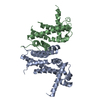
| ||||||||
|---|---|---|---|---|---|---|---|---|---|
| 1 |
| ||||||||
| Unit cell |
|
-Components
| #1: Protein | Mass: 25071.770 Da / Num. of mol.: 2 Source method: isolated from a genetically manipulated source Source: (gene. exp.) Pseudomonas aeruginosa (bacteria) / Strain: UCBPP-PA14 / Gene: PA14_25800 / Production host: #2: Water | ChemComp-HOH / |  Mass: 18.015 Da / Num. of mol.: 255 / Source method: isolated from a natural source / Formula: H2O Mass: 18.015 Da / Num. of mol.: 255 / Source method: isolated from a natural source / Formula: H2O |
|---|
-Experimental details
-Experiment
| Experiment | Method: X-RAY DIFFRACTION / Number of used crystals: 1 |
|---|
- Sample preparation
| Crystal | Density Matthews: 2.35 Å3/Da / Density % sol: 47.75 % |
|---|---|
| Crystal grow | Temperature: 293.15 K / Method: vapor diffusion, sitting drop / Details: PEG 3000, 100mM Tris, 200mM Nacl |
-Data collection
| Diffraction | Mean temperature: 100 K / Serial crystal experiment: N |
|---|---|
| Diffraction source | Source: SEALED TUBE / Type: BRUKER D8 QUEST / Wavelength: 1.5 Å |
| Detector | Type: Bruker PHOTON III / Detector: PIXEL / Date: Oct 10, 2021 |
| Radiation | Protocol: SINGLE WAVELENGTH / Monochromatic (M) / Laue (L): M / Scattering type: x-ray |
| Radiation wavelength | Wavelength: 1.5 Å / Relative weight: 1 |
| Reflection | Resolution: 2.3→31.96 Å / Num. obs: 21122 / % possible obs: 99.2 % / Redundancy: 18.7 % / CC1/2: 0.999 / Net I/σ(I): 27.8 |
| Reflection shell | Resolution: 2.3→2.38 Å / Num. unique obs: 2059 / CC1/2: 0.96 |
- Processing
| Software |
| |||||||||||||||||||||||||||||||||||||||||||||||||||||||||||||||||||||||||||||||||||||||||||||||||||||||||
|---|---|---|---|---|---|---|---|---|---|---|---|---|---|---|---|---|---|---|---|---|---|---|---|---|---|---|---|---|---|---|---|---|---|---|---|---|---|---|---|---|---|---|---|---|---|---|---|---|---|---|---|---|---|---|---|---|---|---|---|---|---|---|---|---|---|---|---|---|---|---|---|---|---|---|---|---|---|---|---|---|---|---|---|---|---|---|---|---|---|---|---|---|---|---|---|---|---|---|---|---|---|---|---|---|---|---|
| Refinement | Method to determine structure: MIRAS / Resolution: 2.303→22.368 Å / SU ML: 0.25 / Cross valid method: FREE R-VALUE / σ(F): 0 / Phase error: 23.49 / Stereochemistry target values: MLHL
| |||||||||||||||||||||||||||||||||||||||||||||||||||||||||||||||||||||||||||||||||||||||||||||||||||||||||
| Solvent computation | Shrinkage radii: 0.9 Å / VDW probe radii: 1.11 Å / Solvent model: FLAT BULK SOLVENT MODEL | |||||||||||||||||||||||||||||||||||||||||||||||||||||||||||||||||||||||||||||||||||||||||||||||||||||||||
| Refinement step | Cycle: LAST / Resolution: 2.303→22.368 Å
| |||||||||||||||||||||||||||||||||||||||||||||||||||||||||||||||||||||||||||||||||||||||||||||||||||||||||
| Refine LS restraints |
| |||||||||||||||||||||||||||||||||||||||||||||||||||||||||||||||||||||||||||||||||||||||||||||||||||||||||
| LS refinement shell |
|
