cytotoxic T cell differentiation / T cell mediated immunity / cell surface receptor signaling pathway / external side of plasma membrane Similarity search - Function
Protocol: SINGLE WAVELENGTH / Monochromatic (M) / Laue (L): M / Scattering type: x-ray
Radiation wavelength
Wavelength: 0.9979 Å / Relative weight: 1
Reflection
Resolution: 1.35→50 Å / Num. obs: 20802 / % possible obs: 99.2 % / Redundancy: 12.4 % / CC1/2: 0.96 / Net I/σ(I): 2.19
Reflection shell
Resolution: 1.35→1.37 Å / CC1/2: 0.96
-
Processing
Software
Name
Version
Classification
REFMAC
5.8.0267
refinement
HKL-3000
datareduction
HKL-3000
datascaling
PHENIX
phasing
Refinement
Method to determine structure: MOLECULAR REPLACEMENT / Resolution: 1.35→35.324 Å / Cor.coef. Fo:Fc: 0.96 / Cor.coef. Fo:Fc free: 0.965 / SU B: 0.006 / SU ML: 0 / Cross valid method: THROUGHOUT / ESU R: 0.063 / ESU R Free: 0.064 Details: Hydrogens have been added in their riding positions
Rfactor
Num. reflection
% reflection
Rfree
0.2161
1113
5.35 %
Rwork
0.2124
19689
-
all
0.213
-
-
obs
-
20802
99.218 %
Solvent computation
Ion probe radii: 0.8 Å / Shrinkage radii: 0.8 Å / VDW probe radii: 1.2 Å / Solvent model: MASK BULK SOLVENT
Displacement parameters
Biso mean: 21.784 Å2
Baniso -1
Baniso -2
Baniso -3
1-
-0.188 Å2
-0 Å2
-0.506 Å2
2-
-
-0.232 Å2
-0 Å2
3-
-
-
0.463 Å2
Refinement step
Cycle: LAST / Resolution: 1.35→35.324 Å
Protein
Nucleic acid
Ligand
Solvent
Total
Num. atoms
786
0
0
125
911
LS refinement shell
Resolution (Å)
Rfactor Rfree
Num. reflection Rfree
Rfactor Rwork
Num. reflection Rwork
Refine-ID
% reflection obs (%)
1.35-1.385
0.4
83
0.433
1416
X-RAY DIFFRACTION
96.0282
1.385-1.423
0.412
93
0.384
1389
X-RAY DIFFRACTION
99.5299
1.423-1.464
0.38
79
0.332
1356
X-RAY DIFFRACTION
99.308
1.464-1.509
0.284
78
0.268
1329
X-RAY DIFFRACTION
99.3644
1.509-1.559
0.341
65
0.257
1302
X-RAY DIFFRACTION
99.7082
1.559-1.613
0.257
62
0.237
1267
X-RAY DIFFRACTION
99.4016
1.613-1.674
0.26
84
0.227
1214
X-RAY DIFFRACTION
99.6163
1.674-1.742
0.25
68
0.222
1146
X-RAY DIFFRACTION
99.5082
1.742-1.819
0.288
46
0.234
1131
X-RAY DIFFRACTION
99.4928
1.819-1.908
0.237
43
0.219
1096
X-RAY DIFFRACTION
99.3025
1.908-2.011
0.248
50
0.216
1027
X-RAY DIFFRACTION
99.5379
2.011-2.132
0.253
50
0.218
952
X-RAY DIFFRACTION
99.3062
2.132-2.279
0.217
79
0.2
885
X-RAY DIFFRACTION
99.6898
2.279-2.461
0.222
40
0.208
860
X-RAY DIFFRACTION
99.5575
2.461-2.694
0.241
33
0.205
791
X-RAY DIFFRACTION
99.6372
2.694-3.01
0.172
36
0.195
722
X-RAY DIFFRACTION
99.6058
3.01-3.472
0.181
43
0.172
620
X-RAY DIFFRACTION
99.5496
3.472-4.243
0.145
46
0.153
514
X-RAY DIFFRACTION
100
4.243-5.959
0.157
21
0.184
435
X-RAY DIFFRACTION
99.7812
5.959-35.324
0.241
14
0.259
237
X-RAY DIFFRACTION
96.5385
+
About Yorodumi
-
News
-
Feb 9, 2022. New format data for meta-information of EMDB entries
New format data for meta-information of EMDB entries
Version 3 of the EMDB header file is now the official format.
The previous official version 1.9 will be removed from the archive.
In the structure databanks used in Yorodumi, some data are registered as the other names, "COVID-19 virus" and "2019-nCoV". Here are the details of the virus and the list of structure data.
Jan 31, 2019. EMDB accession codes are about to change! (news from PDBe EMDB page)
EMDB accession codes are about to change! (news from PDBe EMDB page)
The allocation of 4 digits for EMDB accession codes will soon come to an end. Whilst these codes will remain in use, new EMDB accession codes will include an additional digit and will expand incrementally as the available range of codes is exhausted. The current 4-digit format prefixed with “EMD-” (i.e. EMD-XXXX) will advance to a 5-digit format (i.e. EMD-XXXXX), and so on. It is currently estimated that the 4-digit codes will be depleted around Spring 2019, at which point the 5-digit format will come into force.
The EM Navigator/Yorodumi systems omit the EMD- prefix.
Related info.:Q: What is EMD? / ID/Accession-code notation in Yorodumi/EM Navigator
Yorodumi is a browser for structure data from EMDB, PDB, SASBDB, etc.
This page is also the successor to EM Navigator detail page, and also detail information page/front-end page for Omokage search.
The word "yorodu" (or yorozu) is an old Japanese word meaning "ten thousand". "mi" (miru) is to see.
Related info.:EMDB / PDB / SASBDB / Comparison of 3 databanks / Yorodumi Search / Aug 31, 2016. New EM Navigator & Yorodumi / Yorodumi Papers / Jmol/JSmol / Function and homology information / Changes in new EM Navigator and Yorodumi
 Movie
Movie Controller


 Open data
 Basic information
 Components
 Keywords
 Function and homology information
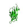
 Scyliorhinus canicula (smaller spotted catshark)
 X-RAY DIFFRACTION /
  Authors
 China, 1items
  Citation
 Structure visualization
 Downloads & links
 Other downloads
 PDBj
PDBj
 Assembly

 Mass: 18.015 Da / Num. of mol.: 125 / Source method: isolated from a natural source / Formula: H2O
 Sample preparation
 Processing