 Movie
Movie Controller

[English] 日本語
 Yorodumi
- PDB-8hwj: Bacterial STING from Epilithonimonas lactis in complex with 3'3'-... -
+ Open data

- Basic information
| Entry | Database: PDB / ID: 8hwj | |||||||||
|---|---|---|---|---|---|---|---|---|---|---|
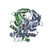
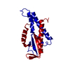
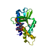
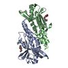
| Title | Bacterial STING from Epilithonimonas lactis in complex with 3'3'-c-di-AMP | |||||||||
 Components Components | CD-NTase-associated protein 12 | |||||||||
 Keywords Keywords | SIGNALING PROTEIN / CD-NTase-associated protein 12 | |||||||||
| Function / homology |  Function and homology information Function and homology informationNAD+ glycohydrolase / NADP+ nucleosidase activity / defense response to virus / nucleotide binding Similarity search - Function | |||||||||
| Biological species |  Epilithonimonas lactis (bacteria) Epilithonimonas lactis (bacteria) | |||||||||
| Method |  X-RAY DIFFRACTION / SYNCHROTRON / MOLECULAR REPLACEMENT / Resolution: 2.553 Å X-RAY DIFFRACTION / SYNCHROTRON / MOLECULAR REPLACEMENT / Resolution: 2.553 Å | |||||||||
 Authors Authors | Wang, Y.-C. / Yang, C.-S. / Hou, M.-H. / Chen, Y. | |||||||||
| Funding support |  Taiwan, 2items Taiwan, 2items
| |||||||||
 Citation Citation | Journal: Nat Commun / Year: 2023 Title: Structural insights into the regulation, ligand recognition, and oligomerization of bacterial STING. Authors: Hou, M.H. / Wang, Y.C. / Yang, C.S. / Liao, K.F. / Chang, J.W. / Shih, O. / Yeh, Y.Q. / Sriramoju, M.K. / Weng, T.W. / Jeng, U.S. / Hsu, S.D. / Chen, Y. | |||||||||
| History |
|
- Structure visualization
| Structure viewer | Molecule: MolmilJmol/JSmol |
|---|
- Downloads & links
-Download
| PDBx/mmCIF format | 8hwj.cif.gz | 146.2 KB | Display | PDBx/mmCIF format |
|---|---|---|---|---|
| PDB format | pdb8hwj.ent.gz | 113.7 KB | Display | PDB format |
| PDBx/mmJSON format | 8hwj.json.gz | Tree view | PDBx/mmJSON format | |
| Others |  Other downloads Other downloads |
-Validation report
| Summary document | 8hwj_validation.pdf.gz | 1.4 MB | Display | wwPDB validaton report |
|---|---|---|---|---|
| Full document | 8hwj_full_validation.pdf.gz | 1.5 MB | Display | |
| Data in XML | 8hwj_validation.xml.gz | 26.4 KB | Display | |
| Data in CIF | 8hwj_validation.cif.gz | 36.6 KB | Display | |
| Arichive directory | https://data.pdbj.org/pub/pdb/validation_reports/hw/8hwjftp://data.pdbj.org/pub/pdb/validation_reports/hw/8hwj | HTTPS FTP |
-Related structure data
-Links
 PDBj
PDBj- Assembly
| Deposited unit | 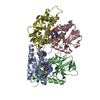
| ||||||||
|---|---|---|---|---|---|---|---|---|---|
| 1 | 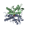
| ||||||||
| 2 | 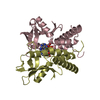
| ||||||||
| Unit cell |
|
-Components
| #1: Protein | Mass: 18813.592 Da / Num. of mol.: 4 Source method: isolated from a genetically manipulated source Details: The GLY4 should be re-indexed as GLY151. / Source: (gene. exp.) Epilithonimonas lactis (bacteria) / Gene: IO89_10965 / Production host: #2: Chemical |   Mass: 658.412 Da / Num. of mol.: 2 / Source method: obtained synthetically / Formula: C20H24N10O12P2 / Feature type: SUBJECT OF INVESTIGATION Mass: 658.412 Da / Num. of mol.: 2 / Source method: obtained synthetically / Formula: C20H24N10O12P2 / Feature type: SUBJECT OF INVESTIGATION#3: Water | ChemComp-HOH / |  Mass: 18.015 Da / Num. of mol.: 172 / Source method: isolated from a natural source / Formula: H2O Mass: 18.015 Da / Num. of mol.: 172 / Source method: isolated from a natural source / Formula: H2OHas ligand of interest | Y | |
|---|
-Experimental details
-Experiment
| Experiment | Method: X-RAY DIFFRACTION / Number of used crystals: 1 |
|---|
- Sample preparation
| Crystal | Density Matthews: 2.22 Å3/Da / Density % sol: 44.61 % |
|---|---|
| Crystal grow | Temperature: 277 K / Method: vapor diffusion, sitting drop Details: 0.1 M MES pH 6.0, 0.1 M Sodium chloride, 30 % v/v PEG 3500 |
-Data collection
| Diffraction | Mean temperature: 100 K / Serial crystal experiment: N |
|---|---|
| Diffraction source | Source: SYNCHROTRON / Site: NSRRC / Beamline: TPS 07A / Wavelength: 0.97626 Å |
| Detector | Type: DECTRIS EIGER X 16M / Detector: PIXEL / Date: Jun 20, 2022 |
| Radiation | Protocol: SINGLE WAVELENGTH / Monochromatic (M) / Laue (L): M / Scattering type: x-ray |
| Radiation wavelength | Wavelength: 0.97626 Å / Relative weight: 1 |
| Reflection | Resolution: 2.55→30 Å / Num. obs: 20886 / % possible obs: 96.5 % / Redundancy: 6.3 % / Rmerge(I) obs: 0.121 / Net I/σ(I): 14.1 |
| Reflection shell | Resolution: 2.55→2.64 Å / Redundancy: 5.6 % / Rmerge(I) obs: 0.578 / Mean I/σ(I) obs: 2.3 / Num. unique obs: 1948 |
- Processing
| Software |
| |||||||||||||||||||||||||||||||||||||||||||||||||||||||||||||||||||||||||||||||||||||||||||||||||||||||||||||||||||||||||||||||||||||||||||||||||||||||||||
|---|---|---|---|---|---|---|---|---|---|---|---|---|---|---|---|---|---|---|---|---|---|---|---|---|---|---|---|---|---|---|---|---|---|---|---|---|---|---|---|---|---|---|---|---|---|---|---|---|---|---|---|---|---|---|---|---|---|---|---|---|---|---|---|---|---|---|---|---|---|---|---|---|---|---|---|---|---|---|---|---|---|---|---|---|---|---|---|---|---|---|---|---|---|---|---|---|---|---|---|---|---|---|---|---|---|---|---|---|---|---|---|---|---|---|---|---|---|---|---|---|---|---|---|---|---|---|---|---|---|---|---|---|---|---|---|---|---|---|---|---|---|---|---|---|---|---|---|---|---|---|---|---|---|---|---|---|
| Refinement | Method to determine structure: MOLECULAR REPLACEMENT / Resolution: 2.553→25.769 Å / Cor.coef. Fo:Fc: 0.939 / Cor.coef. Fo:Fc free: 0.915 / Cross valid method: THROUGHOUT / ESU R Free: 0.33 Details: Hydrogens have been added in their riding positions
| |||||||||||||||||||||||||||||||||||||||||||||||||||||||||||||||||||||||||||||||||||||||||||||||||||||||||||||||||||||||||||||||||||||||||||||||||||||||||||
| Solvent computation | Ion probe radii: 0.8 Å / Shrinkage radii: 0.8 Å / VDW probe radii: 1.2 Å / Solvent model: MASK BULK SOLVENT | |||||||||||||||||||||||||||||||||||||||||||||||||||||||||||||||||||||||||||||||||||||||||||||||||||||||||||||||||||||||||||||||||||||||||||||||||||||||||||
| Displacement parameters | Biso mean: 45.432 Å2
| |||||||||||||||||||||||||||||||||||||||||||||||||||||||||||||||||||||||||||||||||||||||||||||||||||||||||||||||||||||||||||||||||||||||||||||||||||||||||||
| Refinement step | Cycle: LAST / Resolution: 2.553→25.769 Å
| |||||||||||||||||||||||||||||||||||||||||||||||||||||||||||||||||||||||||||||||||||||||||||||||||||||||||||||||||||||||||||||||||||||||||||||||||||||||||||
| Refine LS restraints |
| |||||||||||||||||||||||||||||||||||||||||||||||||||||||||||||||||||||||||||||||||||||||||||||||||||||||||||||||||||||||||||||||||||||||||||||||||||||||||||
| LS refinement shell |
|
