 Movie
Movie Controller

[English] 日本語
 Yorodumi
- PDB-8ht3: Crystal structure of immunoglobulin new antigen receptor variable... -
+ Open data

- Basic information
| Entry | Database: PDB / ID: 8ht3 | ||||||
|---|---|---|---|---|---|---|---|
| Title | Crystal structure of immunoglobulin new antigen receptor variable domain from Okamejei kenojei | ||||||
 Components Components | New Antigen Receptor variable domain | ||||||
 Keywords Keywords |  IMMUNE SYSTEM / Nanobody / Ray vNAR. IMMUNE SYSTEM / Nanobody / Ray vNAR. | ||||||
| Biological species |  Okamejei kenojei (ocellate spot skate) Okamejei kenojei (ocellate spot skate) | ||||||
| Method | X-RAY DIFFRACTION / MOLECULAR REPLACEMENT / Resolution: 2.5 Å | ||||||
 Authors Authors | Wen, J. / Li, P. / Bi, Y. | ||||||
| Funding support |  China, 1items China, 1items
| ||||||
 Citation Citation | Journal: Front Mar Sci / Year: 2023 Title: Identification and characterization of IgNAR and VNAR repertoire from the ocellate spot skate (Okamejei kenojei) Authors: Wen, J. / Gong, J. / Li, P. / Deng, P. / Sun, M. / Wu, Y. / Tian, C. / Wang, H. / Bi, Y. | ||||||
| History |
|
- Structure visualization
| Structure viewer | Molecule:  MolmilJmol/JSmol MolmilJmol/JSmol |
|---|
- Downloads & links
-Download
| PDBx/mmCIF format | 8ht3.cif.gz | 34.5 KB | Display | PDBx/mmCIF format |
|---|---|---|---|---|
| PDB format | pdb8ht3.ent.gz | 21.5 KB | Display | PDB format |
| PDBx/mmJSON format | 8ht3.json.gz | Tree view | PDBx/mmJSON format | |
| Others |  Other downloads Other downloads |
-Validation report
| Arichive directory | https://data.pdbj.org/pub/pdb/validation_reports/ht/8ht3ftp://data.pdbj.org/pub/pdb/validation_reports/ht/8ht3 | HTTPS FTP |
|---|
-Links
 PDBj
PDBj
- Assembly
| Deposited unit | 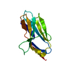
| ||||||||
|---|---|---|---|---|---|---|---|---|---|
| 1 |
| ||||||||
| Unit cell |
|
-Components
| #1: Protein | Mass: 11533.941 Da / Num. of mol.: 1 Source method: isolated from a genetically manipulated source Source: (gene. exp.) Okamejei kenojei (ocellate spot skate) / Production host:  Escherichia coli (E. coli) Escherichia coli (E. coli) |
|---|---|
| #2: Water | ChemComp-HOH / Water Mass: 18.015 Da / Num. of mol.: 53 / Source method: isolated from a natural source / Formula: H2O Mass: 18.015 Da / Num. of mol.: 53 / Source method: isolated from a natural source / Formula: H2O |
-Experimental details
-Experiment
| Experiment | Method: X-RAY DIFFRACTION / Number of used crystals: 1 |
|---|
- Sample preparation
| Crystal | Density Matthews: 2.32 Å3/Da / Density % sol: 47 % |
|---|---|
| Crystal grow | Temperature: 277.15 K / Method: vapor diffusion, sitting drop Details: 0.2 M Lithium sulfate monohydrate/0.1 M Tris pH 8.0/26% w/v PEG4000 |
-Data collection
| Diffraction | Mean temperature: 100 K / Serial crystal experiment: N |
|---|---|
| Diffraction source | Source: ROTATING ANODE / Type: RIGAKU FR-E DW / Wavelength: 1.54184 Å |
| Detector | Type: RIGAKU HyPix-6000HE / Detector: PIXEL / Date: Dec 14, 2022 |
| Radiation | Monochromator: mirror / Protocol: SINGLE WAVELENGTH / Monochromatic (M) / Laue (L): M / Scattering type: x-ray |
| Radiation wavelength | Wavelength: 1.54184 Å / Relative weight: 1 |
| Reflection | Resolution: 2.5→14.05 Å / Num. obs: 4028 / % possible obs: 98.92 % / Redundancy: 5.2 % / CC1/2: 1 / Rmerge(I) obs: 0.02776 / Rpim(I) all: 0.01293 / Rrim(I) all: 0.03077 / Net I/σ(I): 61.28 |
| Reflection shell | Resolution: 2.5→2.589 Å / Num. unique obs: 1866 / CC1/2: 0.997 / % possible all: 97.92 |
- Processing
| Software |
| ||||||||||||||||||||||||||||
|---|---|---|---|---|---|---|---|---|---|---|---|---|---|---|---|---|---|---|---|---|---|---|---|---|---|---|---|---|---|
| Refinement | Method to determine structure: MOLECULAR REPLACEMENT / Resolution: 2.5→14.05 Å / SU ML: 0.31 / Cross valid method: NONE / σ(F): 0 / Phase error: 22.03 / Stereochemistry target values: ML
| ||||||||||||||||||||||||||||
| Solvent computation | Shrinkage radii: 0.9 Å / VDW probe radii: 1.11 Å / Solvent model: FLAT BULK SOLVENT MODEL | ||||||||||||||||||||||||||||
| Refinement step | Cycle: LAST / Resolution: 2.5→14.05 Å
| ||||||||||||||||||||||||||||
| Refine LS restraints |
| ||||||||||||||||||||||||||||
| LS refinement shell |
|
