 Movie
Movie Controller

[English] 日本語
 Yorodumi
- PDB-8hs7: Crystal structure of Vesicle-associated membrane protein-associat... -
+ Open data

- Basic information
| Entry | Database: PDB / ID: 8hs7 | ||||||
|---|---|---|---|---|---|---|---|
| Title | Crystal structure of Vesicle-associated membrane protein-associated protein SCS2 from yeast | ||||||
 Components Components | Vesicle-associated membrane protein-associated protein SCS2 | ||||||
 Keywords Keywords | CELL ADHESION | ||||||
| Function / homology |  Function and homology information Function and homology informationendoplasmic reticulum polarization / regulation of intracellular lipid transport / FFAT motif binding / : / RHOC GTPase cycle / endoplasmic reticulum membrane organization / endoplasmic reticulum inheritance / regulation of phosphatidylinositol dephosphorylation / nucleus-vacuole junction / Insertion of tail-anchored proteins into the endoplasmic reticulum membrane ...endoplasmic reticulum polarization / regulation of intracellular lipid transport / FFAT motif binding / : / RHOC GTPase cycle / endoplasmic reticulum membrane organization / endoplasmic reticulum inheritance / regulation of phosphatidylinositol dephosphorylation / nucleus-vacuole junction / Insertion of tail-anchored proteins into the endoplasmic reticulum membrane / cellular bud tip / phospholipid biosynthetic process / cellular bud neck / reticulophagy / negative regulation of protein import into nucleus / subtelomeric heterochromatin formation / protein-membrane adaptor activity / Neutrophil degranulation / phosphatidylinositol binding / nuclear envelope / nuclear membrane / chromosome, telomeric region / endoplasmic reticulum membrane / endoplasmic reticulum / plasma membrane Similarity search - Function | ||||||
| Biological species |  | ||||||
| Method |  X-RAY DIFFRACTION / SYNCHROTRON / MOLECULAR REPLACEMENT / Resolution: 2 Å X-RAY DIFFRACTION / SYNCHROTRON / MOLECULAR REPLACEMENT / Resolution: 2 Å | ||||||
 Authors Authors | Xu, T. | ||||||
| Funding support |  China, 1items China, 1items
| ||||||
 Citation Citation | Journal: To Be Published Title: Crystal structure of the major sperm protein domain of SCS2 from saccharomyces cerevisiae Authors: Xu, T. | ||||||
| History |
|
- Structure visualization
| Structure viewer | Molecule: MolmilJmol/JSmol |
|---|
- Downloads & links
-Download
| PDBx/mmCIF format | 8hs7.cif.gz | 67.5 KB | Display | PDBx/mmCIF format |
|---|---|---|---|---|
| PDB format | pdb8hs7.ent.gz | 46.1 KB | Display | PDB format |
| PDBx/mmJSON format | 8hs7.json.gz | Tree view | PDBx/mmJSON format | |
| Others |  Other downloads Other downloads |
-Validation report
| Arichive directory | https://data.pdbj.org/pub/pdb/validation_reports/hs/8hs7ftp://data.pdbj.org/pub/pdb/validation_reports/hs/8hs7 | HTTPS FTP |
|---|
-Related structure data
| Similar structure data |
|---|
-Links
 PDBj
PDBj


- Assembly
| Deposited unit | 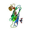
| ||||||||||||
|---|---|---|---|---|---|---|---|---|---|---|---|---|---|
| 1 |
| ||||||||||||
| Unit cell |
| ||||||||||||
| Components on special symmetry positions |
|
-Components
| #1: Protein | Mass: 13932.947 Da / Num. of mol.: 1 / Fragment: MSP domain Source method: isolated from a genetically manipulated source Source: (gene. exp.) Gene: SCS2, YER120W / Production host:  |
|---|---|
| #2: Chemical | ChemComp-EDT / {[-(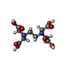  Mass: 292.243 Da / Num. of mol.: 1 / Source method: obtained synthetically / Formula: C10H16N2O8 Mass: 292.243 Da / Num. of mol.: 1 / Source method: obtained synthetically / Formula: C10H16N2O8 |
| #3: Water | ChemComp-HOH /  Mass: 18.015 Da / Num. of mol.: 47 / Source method: isolated from a natural source / Formula: H2O Mass: 18.015 Da / Num. of mol.: 47 / Source method: isolated from a natural source / Formula: H2O |
| Has ligand of interest | N |
-Experimental details
-Experiment
| Experiment | Method: X-RAY DIFFRACTION / Number of used crystals: 1 |
|---|
- Sample preparation
| Crystal | Density Matthews: 2.76 Å3/Da / Density % sol: 55.46 % |
|---|---|
| Crystal grow | Temperature: 289.15 K / Method: vapor diffusion, sitting drop / Details: 25% PEG 3350, 0.1 mM tris-bis (pH 5.5). 0.1 M EDTA |
-Data collection
| Diffraction | Mean temperature: 100 K / Serial crystal experiment: N |
|---|---|
| Diffraction source | Source: SYNCHROTRON / Site: SSRF / Beamline: BL19U1 / Wavelength: 0.97852 Å |
| Detector | Type: DECTRIS PILATUS 6M / Detector: PIXEL / Date: May 29, 2020 |
| Radiation | Protocol: SINGLE WAVELENGTH / Monochromatic (M) / Laue (L): M / Scattering type: x-ray |
| Radiation wavelength | Wavelength: 0.97852 Å / Relative weight: 1 |
| Reflection | Resolution: 2→50 Å / Num. obs: 10680 / % possible obs: 99.81 % / Redundancy: 19 % / Biso Wilson estimate: 39.82 Å2 / CC1/2: 1 / Rmerge(I) obs: 0.05229 / Rpim(I) all: 0.01233 / Net I/σ(I): 37.12 |
| Reflection shell | Resolution: 2→2.09 Å / Rmerge(I) obs: 0.6431 / Mean I/σ(I) obs: 4.49 / Num. unique obs: 1312 / CC1/2: 0.95 / Rpim(I) all: 0.1588 |
- Processing
| Software |
| |||||||||||||||||||||||||||||||||||||||||||||||||||||||||||||||
|---|---|---|---|---|---|---|---|---|---|---|---|---|---|---|---|---|---|---|---|---|---|---|---|---|---|---|---|---|---|---|---|---|---|---|---|---|---|---|---|---|---|---|---|---|---|---|---|---|---|---|---|---|---|---|---|---|---|---|---|---|---|---|---|---|
| Refinement | Method to determine structure: MOLECULAR REPLACEMENT / Resolution: 2→40.92 Å / SU ML: 0.2474 / Cross valid method: FREE R-VALUE / σ(F): 1.35 / Phase error: 29.9488 Stereochemistry target values: GeoStd + Monomer Library + CDL v1.2
| |||||||||||||||||||||||||||||||||||||||||||||||||||||||||||||||
| Solvent computation | Shrinkage radii: 0.9 Å / VDW probe radii: 1.1 Å / Solvent model: FLAT BULK SOLVENT MODEL | |||||||||||||||||||||||||||||||||||||||||||||||||||||||||||||||
| Displacement parameters | Biso mean: 45.45 Å2 | |||||||||||||||||||||||||||||||||||||||||||||||||||||||||||||||
| Refinement step | Cycle: LAST / Resolution: 2→40.92 Å
| |||||||||||||||||||||||||||||||||||||||||||||||||||||||||||||||
| Refine LS restraints |
| |||||||||||||||||||||||||||||||||||||||||||||||||||||||||||||||
| LS refinement shell |
|
