 Movie
Movie Controller

[English] 日本語
 Yorodumi
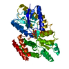
- PDB-8hqq: Crystal structure of the glucose-binding protein SAR11_0769 from ... -
+ Open data

- Basic information
| Entry | Database: PDB / ID: 8hqq | ||||||
|---|---|---|---|---|---|---|---|
| Title | Crystal structure of the glucose-binding protein SAR11_0769 from "Candidatus Pelagibacter ubique" HTCC1062 bound to glucose | ||||||
 Components Components | Probable binding protein component of ABC sugar transporter | ||||||
 Keywords Keywords | TRANSPORT PROTEIN / ABC transporter / glucose transporter / periplasmic binding protein / solute-binding protein | ||||||
| Function / homology | : / Bacterial extracellular solute-binding protein / Bacterial extracellular solute-binding protein / periplasmic space / beta-D-glucopyranose / Probable sugar-binding periplasmic protein Function and homology information Function and homology information | ||||||
| Biological species |  Candidatus Pelagibacter ubique HTCC1062 (bacteria) Candidatus Pelagibacter ubique HTCC1062 (bacteria) | ||||||
| Method |  X-RAY DIFFRACTION / SYNCHROTRON / MOLECULAR REPLACEMENT / Resolution: 1.86 Å X-RAY DIFFRACTION / SYNCHROTRON / MOLECULAR REPLACEMENT / Resolution: 1.86 Å | ||||||
 Authors Authors | Clifton, B.E. / Laurino, P. | ||||||
| Funding support |  Japan, 1items Japan, 1items
| ||||||
 Citation Citation | Journal: Nature / Year: 2024 Title: The ultra-high affinity transport proteins of ubiquitous marine bacteria. Authors: Clifton, B.E. / Alcolombri, U. / Uechi, G.I. / Jackson, C.J. / Laurino, P. | ||||||
| History |
|
- Structure visualization
| Structure viewer | Molecule: MolmilJmol/JSmol |
|---|
- Downloads & links
-Download
| PDBx/mmCIF format | 8hqq.cif.gz | 164 KB | Display | PDBx/mmCIF format |
|---|---|---|---|---|
| PDB format | pdb8hqq.ent.gz | 127.8 KB | Display | PDB format |
| PDBx/mmJSON format | 8hqq.json.gz | Tree view | PDBx/mmJSON format | |
| Others |  Other downloads Other downloads |
-Validation report
| Arichive directory | https://data.pdbj.org/pub/pdb/validation_reports/hq/8hqqftp://data.pdbj.org/pub/pdb/validation_reports/hq/8hqq | HTTPS FTP |
|---|
-Related structure data

-Links
 PDBj
PDBj


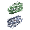
- Assembly
| Deposited unit | 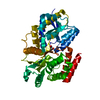
| ||||||||
|---|---|---|---|---|---|---|---|---|---|
| 1 |
| ||||||||
| Unit cell |
|
-Components
| #1: Protein | Mass: 44926.027 Da / Num. of mol.: 1 Source method: isolated from a genetically manipulated source Source: (gene. exp.) Candidatus Pelagibacter ubique HTCC1062 (bacteria)Gene: SAR11_0769 / Plasmid: pET-28a(+) / Production host: |
|---|---|
| #2: Sugar | ChemComp-BGC /   Type: D-saccharide, beta linking / Mass: 180.156 Da / Num. of mol.: 1 / Source method: obtained synthetically / Formula: C6H12O6 / Feature type: SUBJECT OF INVESTIGATION Type: D-saccharide, beta linking / Mass: 180.156 Da / Num. of mol.: 1 / Source method: obtained synthetically / Formula: C6H12O6 / Feature type: SUBJECT OF INVESTIGATION |
| #3: Water | ChemComp-HOH /  Mass: 18.015 Da / Num. of mol.: 99 / Source method: isolated from a natural source / Formula: H2O Mass: 18.015 Da / Num. of mol.: 99 / Source method: isolated from a natural source / Formula: H2O |
| Has ligand of interest | Y |
| Has protein modification | Y |
-Experimental details
-Experiment
| Experiment | Method: X-RAY DIFFRACTION / Number of used crystals: 1 |
|---|
- Sample preparation
| Crystal | Density Matthews: 2.11 Å3/Da / Density % sol: 41.63 % |
|---|---|
| Crystal grow | Temperature: 293 K / Method: vapor diffusion, hanging drop / pH: 6.5 Details: 1 uL 12 mg/mL protein + 1 uL 18.5% (w/v) PEG 1500, 10% (v/v) isopropanol, 0.1 M Bis-Tris pH 6.5. Crystal was cryoprotected in 15% (w/v) PEG 1500, 30% (v/v) ethylene glycol, 10% (v/v) ...Details: 1 uL 12 mg/mL protein + 1 uL 18.5% (w/v) PEG 1500, 10% (v/v) isopropanol, 0.1 M Bis-Tris pH 6.5. Crystal was cryoprotected in 15% (w/v) PEG 1500, 30% (v/v) ethylene glycol, 10% (v/v) isopropanol, 0.1 M Bis-Tris pH 6.5. |
-Data collection
| Diffraction | Mean temperature: 100 K / Serial crystal experiment: N |
|---|---|
| Diffraction source | Source: SYNCHROTRON / Site: SPring-8 / Beamline: BL32XU / Wavelength: 1 Å |
| Detector | Type: DECTRIS EIGER X 9M / Detector: PIXEL / Date: Oct 25, 2022 |
| Radiation | Protocol: SINGLE WAVELENGTH / Monochromatic (M) / Laue (L): M / Scattering type: x-ray |
| Radiation wavelength | Wavelength: 1 Å / Relative weight: 1 |
| Reflection | Resolution: 1.86→47.02 Å / Num. obs: 33089 / % possible obs: 99 % / Redundancy: 12 % / CC1/2: 0.986 / Rmerge(I) obs: 0.239 / Rrim(I) all: 0.248 / Net I/σ(I): 6.28 |
| Reflection shell | Resolution: 1.86→1.98 Å / Redundancy: 9.49 % / Rmerge(I) obs: 0.521 / Mean I/σ(I) obs: 1.08 / Num. unique obs: 4941 / CC1/2: 0.913 / Rrim(I) all: 0.545 / % possible all: 94.4 |
- Processing
| Software |
| |||||||||||||||||||||||||||||||||||||||||||||||||||||||||||||||||||||||||||||||||||||||||||||||||||||||||||||||||||||||||||||||||||||||||||||||||||||||||||||||||||||||||||||||||||||||||||||||||||||||||||||||||||||||||||||||||
|---|---|---|---|---|---|---|---|---|---|---|---|---|---|---|---|---|---|---|---|---|---|---|---|---|---|---|---|---|---|---|---|---|---|---|---|---|---|---|---|---|---|---|---|---|---|---|---|---|---|---|---|---|---|---|---|---|---|---|---|---|---|---|---|---|---|---|---|---|---|---|---|---|---|---|---|---|---|---|---|---|---|---|---|---|---|---|---|---|---|---|---|---|---|---|---|---|---|---|---|---|---|---|---|---|---|---|---|---|---|---|---|---|---|---|---|---|---|---|---|---|---|---|---|---|---|---|---|---|---|---|---|---|---|---|---|---|---|---|---|---|---|---|---|---|---|---|---|---|---|---|---|---|---|---|---|---|---|---|---|---|---|---|---|---|---|---|---|---|---|---|---|---|---|---|---|---|---|---|---|---|---|---|---|---|---|---|---|---|---|---|---|---|---|---|---|---|---|---|---|---|---|---|---|---|---|---|---|---|---|---|---|---|---|---|---|---|---|---|---|---|---|---|---|---|---|---|
| Refinement | Method to determine structure: MOLECULAR REPLACEMENT Starting model: AlphaFold Resolution: 1.86→38.4 Å / SU ML: 0.27 / Cross valid method: FREE R-VALUE / σ(F): 1.35 / Phase error: 27.36 / Stereochemistry target values: ML Details: Initial refinement by simulated annealing in Phenix, followed by iterative real-space and reciprocal-space refinement in COOT and Refmac. Final rounds of reciprocal-space refinement ...Details: Initial refinement by simulated annealing in Phenix, followed by iterative real-space and reciprocal-space refinement in COOT and Refmac. Final rounds of reciprocal-space refinement including TLS refinement were performed in Phenix.
| |||||||||||||||||||||||||||||||||||||||||||||||||||||||||||||||||||||||||||||||||||||||||||||||||||||||||||||||||||||||||||||||||||||||||||||||||||||||||||||||||||||||||||||||||||||||||||||||||||||||||||||||||||||||||||||||||
| Solvent computation | Shrinkage radii: 0.9 Å / VDW probe radii: 1.1 Å / Solvent model: FLAT BULK SOLVENT MODEL | |||||||||||||||||||||||||||||||||||||||||||||||||||||||||||||||||||||||||||||||||||||||||||||||||||||||||||||||||||||||||||||||||||||||||||||||||||||||||||||||||||||||||||||||||||||||||||||||||||||||||||||||||||||||||||||||||
| Refinement step | Cycle: LAST / Resolution: 1.86→38.4 Å
| |||||||||||||||||||||||||||||||||||||||||||||||||||||||||||||||||||||||||||||||||||||||||||||||||||||||||||||||||||||||||||||||||||||||||||||||||||||||||||||||||||||||||||||||||||||||||||||||||||||||||||||||||||||||||||||||||
| Refine LS restraints |
| |||||||||||||||||||||||||||||||||||||||||||||||||||||||||||||||||||||||||||||||||||||||||||||||||||||||||||||||||||||||||||||||||||||||||||||||||||||||||||||||||||||||||||||||||||||||||||||||||||||||||||||||||||||||||||||||||
| LS refinement shell |
| |||||||||||||||||||||||||||||||||||||||||||||||||||||||||||||||||||||||||||||||||||||||||||||||||||||||||||||||||||||||||||||||||||||||||||||||||||||||||||||||||||||||||||||||||||||||||||||||||||||||||||||||||||||||||||||||||
| Refinement TLS params. | Method: refined / Refine-ID: X-RAY DIFFRACTION
| |||||||||||||||||||||||||||||||||||||||||||||||||||||||||||||||||||||||||||||||||||||||||||||||||||||||||||||||||||||||||||||||||||||||||||||||||||||||||||||||||||||||||||||||||||||||||||||||||||||||||||||||||||||||||||||||||
| Refinement TLS group |
|
