 Movie
Movie Controller

[English] 日本語
 Yorodumi
- PDB-8hqa: Crystal structure of the ectodomain of the MlaD protein from Esch... -
+ Open data

- Basic information
| Entry | Database: PDB / ID: 8hqa | ||||||
|---|---|---|---|---|---|---|---|
| Title | Crystal structure of the ectodomain of the MlaD protein from Escherichia coli in the resting state | ||||||
 Components Components | Intermembrane phospholipid transport system binding protein MlaD | ||||||
 Keywords Keywords | TRANSPORT PROTEIN / ABC transporter / Asymmetric protomer movement / Central channel / Gram-negative bacteria / Mla system / Transport | ||||||
| Function / homology |  Function and homology information Function and homology informationphospholipid transfer activity / intermembrane phospholipid transfer / phospholipid-translocating ATPase complex / phospholipid transport / phospholipid binding / membrane / plasma membrane Similarity search - Function | ||||||
| Biological species |  | ||||||
| Method |  X-RAY DIFFRACTION / MOLECULAR REPLACEMENT / Resolution: 3.2 Å X-RAY DIFFRACTION / MOLECULAR REPLACEMENT / Resolution: 3.2 Å | ||||||
 Authors Authors | Dutta, A. / Kanaujia, S.P. | ||||||
| Funding support |  India, 1items India, 1items
| ||||||
 Citation Citation | Journal: Protein J. / Year: 2024 Title: The Structural Features of MlaD Illuminate its Unique Ligand-Transporting Mechanism and Ancestry. Authors: Dutta, A. / Kanaujia, S.P. | ||||||
| History |
|
- Structure visualization
| Structure viewer | Molecule: MolmilJmol/JSmol |
|---|
- Downloads & links
-Download
| PDBx/mmCIF format | 8hqa.cif.gz | 149.2 KB | Display | PDBx/mmCIF format |
|---|---|---|---|---|
| PDB format | pdb8hqa.ent.gz | 117.5 KB | Display | PDB format |
| PDBx/mmJSON format | 8hqa.json.gz | Tree view | PDBx/mmJSON format | |
| Others |  Other downloads Other downloads |
-Validation report
| Summary document | 8hqa_validation.pdf.gz | 450.2 KB | Display | wwPDB validaton report |
|---|---|---|---|---|
| Full document | 8hqa_full_validation.pdf.gz | 455.2 KB | Display | |
| Data in XML | 8hqa_validation.xml.gz | 14.3 KB | Display | |
| Data in CIF | 8hqa_validation.cif.gz | 18.9 KB | Display | |
| Arichive directory | https://data.pdbj.org/pub/pdb/validation_reports/hq/8hqaftp://data.pdbj.org/pub/pdb/validation_reports/hq/8hqa | HTTPS FTP |
-Related structure data


-Links
 PDBj
PDBj- Assembly
| Deposited unit | 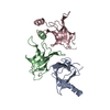
| ||||||||||||
|---|---|---|---|---|---|---|---|---|---|---|---|---|---|
| 1 | 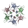
| ||||||||||||
| Unit cell |
| ||||||||||||
| Noncrystallographic symmetry (NCS) | NCS domain:
|
-Components
| #1: Protein | Mass: 17462.348 Da / Num. of mol.: 3 Source method: isolated from a genetically manipulated source Source: (gene. exp.) #2: Water | ChemComp-HOH / |  Mass: 18.015 Da / Num. of mol.: 22 / Source method: isolated from a natural source / Formula: H2O Mass: 18.015 Da / Num. of mol.: 22 / Source method: isolated from a natural source / Formula: H2O |
|---|
-Experimental details
-Experiment
| Experiment | Method: X-RAY DIFFRACTION / Number of used crystals: 1 |
|---|
- Sample preparation
| Crystal | Density Matthews: 2.14 Å3/Da / Density % sol: 42.64 % / Description: Tetragonal |
|---|---|
| Crystal grow | Temperature: 293 K / Method: microbatch / pH: 8.5 Details: 0.01 M nickel(II) chloride hexahydrate, 0.1 M tris pH 8.5, 20% (w/v) PEG monoethyl ether 2000 |
-Data collection
| Diffraction | Mean temperature: 100 K / Serial crystal experiment: N |
|---|---|
| Diffraction source | Source: ROTATING ANODE / Type: RIGAKU MICROMAX-007 HF / Wavelength: 1.5418 Å |
| Detector | Type: RIGAKU RAXIS IV++ / Detector: IMAGE PLATE / Date: Dec 12, 2019 / Details: VariMax HF |
| Radiation | Protocol: SINGLE WAVELENGTH / Monochromatic (M) / Laue (L): M / Scattering type: x-ray |
| Radiation wavelength | Wavelength: 1.5418 Å / Relative weight: 1 |
| Reflection | Resolution: 3.2→77.02 Å / Num. obs: 8680 / % possible obs: 99.8 % / Redundancy: 10 % / CC1/2: 0.994 / Rmerge(I) obs: 0.166 / Rpim(I) all: 0.053 / Rrim(I) all: 0.175 / Χ2: 0.92 / Net I/σ(I): 9.6 / Num. measured all: 86434 |
| Reflection shell | Resolution: 3.2→3.42 Å / % possible obs: 100 % / Redundancy: 10.2 % / Rmerge(I) obs: 0.552 / Num. measured all: 15458 / Num. unique obs: 1516 / CC1/2: 0.934 / Rpim(I) all: 0.177 / Rrim(I) all: 0.582 / Χ2: 0.72 / Net I/σ(I) obs: 3.5 |
- Processing
| Software |
| ||||||||||||||||||||||||||||||||||||||||||||||||||||||||||||||||||||||||||||||||||||||||||||||||||||||||||||||||||||||||||||||||||||||||||||||||||||||||||||||||||||||||||||||||||||||
|---|---|---|---|---|---|---|---|---|---|---|---|---|---|---|---|---|---|---|---|---|---|---|---|---|---|---|---|---|---|---|---|---|---|---|---|---|---|---|---|---|---|---|---|---|---|---|---|---|---|---|---|---|---|---|---|---|---|---|---|---|---|---|---|---|---|---|---|---|---|---|---|---|---|---|---|---|---|---|---|---|---|---|---|---|---|---|---|---|---|---|---|---|---|---|---|---|---|---|---|---|---|---|---|---|---|---|---|---|---|---|---|---|---|---|---|---|---|---|---|---|---|---|---|---|---|---|---|---|---|---|---|---|---|---|---|---|---|---|---|---|---|---|---|---|---|---|---|---|---|---|---|---|---|---|---|---|---|---|---|---|---|---|---|---|---|---|---|---|---|---|---|---|---|---|---|---|---|---|---|---|---|---|---|
| Refinement | Method to determine structure: MOLECULAR REPLACEMENT / Resolution: 3.2→61.93 Å / Cor.coef. Fo:Fc: 0.913 / Cor.coef. Fo:Fc free: 0.9 / SU B: 48.063 / SU ML: 0.381 / Cross valid method: THROUGHOUT / ESU R Free: 0.513 / Stereochemistry target values: MAXIMUM LIKELIHOOD / Details: HYDROGENS HAVE BEEN ADDED IN THE RIDING POSITIONS
| ||||||||||||||||||||||||||||||||||||||||||||||||||||||||||||||||||||||||||||||||||||||||||||||||||||||||||||||||||||||||||||||||||||||||||||||||||||||||||||||||||||||||||||||||||||||
| Solvent computation | Ion probe radii: 0.8 Å / Shrinkage radii: 0.8 Å / VDW probe radii: 1.2 Å / Solvent model: MASK | ||||||||||||||||||||||||||||||||||||||||||||||||||||||||||||||||||||||||||||||||||||||||||||||||||||||||||||||||||||||||||||||||||||||||||||||||||||||||||||||||||||||||||||||||||||||
| Displacement parameters | Biso mean: 70.249 Å2
| ||||||||||||||||||||||||||||||||||||||||||||||||||||||||||||||||||||||||||||||||||||||||||||||||||||||||||||||||||||||||||||||||||||||||||||||||||||||||||||||||||||||||||||||||||||||
| Refinement step | Cycle: 1 / Resolution: 3.2→61.93 Å
| ||||||||||||||||||||||||||||||||||||||||||||||||||||||||||||||||||||||||||||||||||||||||||||||||||||||||||||||||||||||||||||||||||||||||||||||||||||||||||||||||||||||||||||||||||||||
| Refine LS restraints |
|