 Movie
Movie Controller

[English] 日本語
 Yorodumi
- PDB-8hp3: Crystal structure of meso-diaminopimelate dehydrogenase from Prev... -
+ Open data

- Basic information
| Entry | Database: PDB / ID: 8hp3 | ||||||
|---|---|---|---|---|---|---|---|
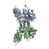
| Title | Crystal structure of meso-diaminopimelate dehydrogenase from Prevotella timonensis | ||||||
 Components Components | Meso-diaminopimelate D-dehydrogenase | ||||||
 Keywords Keywords | OXIDOREDUCTASE / C2221 / fold | ||||||
| Function / homology |  Function and homology information Function and homology informationdiaminopimelate dehydrogenase / diaminopimelate dehydrogenase activity / diaminopimelate biosynthetic process / lysine biosynthetic process via diaminopimelate / nucleotide binding Similarity search - Function | ||||||
| Biological species |  Prevotella timonensis (bacteria) Prevotella timonensis (bacteria) | ||||||
| Method |  X-RAY DIFFRACTION / MOLECULAR REPLACEMENT / Resolution: 3.07 Å X-RAY DIFFRACTION / MOLECULAR REPLACEMENT / Resolution: 3.07 Å | ||||||
 Authors Authors | Tan, Y. / Song, W. | ||||||
| Funding support | 1items
| ||||||
 Citation Citation | Journal: Appl.Environ.Microbiol. / Year: 2023 Title: Rational Design of Meso -Diaminopimelate Dehydrogenase with Enhanced Reductive Amination Activity for Efficient Production of d- p -Hydroxyphenylglycine. Authors: Tan, Y. / Gao, C. / Song, W. / Wei, W. / Liu, J. / Gao, C. / Guo, L. / Chen, X. / Liu, L. / Wu, J. | ||||||
| History |
|
- Structure visualization
| Structure viewer | Molecule: MolmilJmol/JSmol |
|---|
- Downloads & links
-Download
| PDBx/mmCIF format | 8hp3.cif.gz | 183.4 KB | Display | PDBx/mmCIF format |
|---|---|---|---|---|
| PDB format | pdb8hp3.ent.gz | 146.7 KB | Display | PDB format |
| PDBx/mmJSON format | 8hp3.json.gz | Tree view | PDBx/mmJSON format | |
| Others |  Other downloads Other downloads |
-Validation report
| Arichive directory | https://data.pdbj.org/pub/pdb/validation_reports/hp/8hp3ftp://data.pdbj.org/pub/pdb/validation_reports/hp/8hp3 | HTTPS FTP |
|---|
-Related structure data
-Links
 PDBj
PDBj

- Assembly
| Deposited unit | 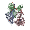
| ||||||||||||
|---|---|---|---|---|---|---|---|---|---|---|---|---|---|
| 1 | 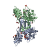
| ||||||||||||
| 2 | 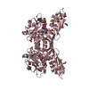
| ||||||||||||
| Unit cell |
| ||||||||||||
| Noncrystallographic symmetry (NCS) | NCS domain:
|
-Components
| #1: Protein | Mass: 32584.482 Da / Num. of mol.: 3 Source method: isolated from a genetically manipulated source Source: (gene. exp.) Prevotella timonensis (bacteria) / Gene: BFS16_11360 / Production host: References: UniProt: A0A2K0XCZ3, diaminopimelate dehydrogenase #2: Chemical |   Mass: 745.421 Da / Num. of mol.: 3 / Source method: obtained synthetically / Formula: C21H30N7O17P3 / Feature type: SUBJECT OF INVESTIGATION Mass: 745.421 Da / Num. of mol.: 3 / Source method: obtained synthetically / Formula: C21H30N7O17P3 / Feature type: SUBJECT OF INVESTIGATION#3: Chemical | ChemComp-SO4 /   Mass: 96.063 Da / Num. of mol.: 8 / Source method: obtained synthetically / Formula: SO4 / Feature type: SUBJECT OF INVESTIGATION Mass: 96.063 Da / Num. of mol.: 8 / Source method: obtained synthetically / Formula: SO4 / Feature type: SUBJECT OF INVESTIGATION#4: Chemical | ChemComp-EDO /   Mass: 62.068 Da / Num. of mol.: 9 / Source method: obtained synthetically / Formula: C2H6O2 / Feature type: SUBJECT OF INVESTIGATION Mass: 62.068 Da / Num. of mol.: 9 / Source method: obtained synthetically / Formula: C2H6O2 / Feature type: SUBJECT OF INVESTIGATION#5: Water | ChemComp-HOH / |  Mass: 18.015 Da / Num. of mol.: 30 / Source method: isolated from a natural source / Formula: H2O Mass: 18.015 Da / Num. of mol.: 30 / Source method: isolated from a natural source / Formula: H2OHas ligand of interest | Y | |
|---|
-Experimental details
-Experiment
| Experiment | Method: X-RAY DIFFRACTION / Number of used crystals: 1 |
|---|
- Sample preparation
| Crystal | Density Matthews: 2.51 Å3/Da / Density % sol: 51.02 % |
|---|---|
| Crystal grow | Temperature: 298 K / Method: vapor diffusion, sitting drop / Details: 0.1 M HEPES, pH 7.5, 0.1 M ammonium sulfate |
-Data collection
| Diffraction | Mean temperature: 100 K / Serial crystal experiment: N |
|---|---|
| Diffraction source | Source: SEALED TUBE / Type: BRUKER D8 QUEST / Wavelength: 1.54178 Å |
| Detector | Type: Bruker PHOTON II / Detector: PIXEL / Date: Nov 25, 2021 |
| Radiation | Protocol: SINGLE WAVELENGTH / Monochromatic (M) / Laue (L): M / Scattering type: x-ray |
| Radiation wavelength | Wavelength: 1.54178 Å / Relative weight: 1 |
| Reflection | Resolution: 3.07→47.53 Å / Num. obs: 18754 / % possible obs: 99.8 % / Redundancy: 4.7 % / Rmerge(I) obs: 0.173 / Net I/σ(I): 7.8 |
| Reflection shell | Resolution: 3.07→3.28 Å / Rmerge(I) obs: 0.66 / Num. unique obs: 3351 |
- Processing
| Software |
| ||||||||||||||||||||||||||||||||||||||||||||||||||||||||||||||||||||||||||||||||||||||||||||||||||||||||||||||||||||||||||||||||||||||||||||||||||||||||||||||||||||||||||||||||||||||
|---|---|---|---|---|---|---|---|---|---|---|---|---|---|---|---|---|---|---|---|---|---|---|---|---|---|---|---|---|---|---|---|---|---|---|---|---|---|---|---|---|---|---|---|---|---|---|---|---|---|---|---|---|---|---|---|---|---|---|---|---|---|---|---|---|---|---|---|---|---|---|---|---|---|---|---|---|---|---|---|---|---|---|---|---|---|---|---|---|---|---|---|---|---|---|---|---|---|---|---|---|---|---|---|---|---|---|---|---|---|---|---|---|---|---|---|---|---|---|---|---|---|---|---|---|---|---|---|---|---|---|---|---|---|---|---|---|---|---|---|---|---|---|---|---|---|---|---|---|---|---|---|---|---|---|---|---|---|---|---|---|---|---|---|---|---|---|---|---|---|---|---|---|---|---|---|---|---|---|---|---|---|---|---|
| Refinement | Method to determine structure: MOLECULAR REPLACEMENT / Resolution: 3.07→47.53 Å / Cor.coef. Fo:Fc: 0.929 / Cor.coef. Fo:Fc free: 0.869 / SU B: 24.307 / SU ML: 0.399 / Cross valid method: THROUGHOUT / ESU R Free: 0.511 / Stereochemistry target values: MAXIMUM LIKELIHOOD / Details: HYDROGENS HAVE BEEN ADDED IN THE RIDING POSITIONS
| ||||||||||||||||||||||||||||||||||||||||||||||||||||||||||||||||||||||||||||||||||||||||||||||||||||||||||||||||||||||||||||||||||||||||||||||||||||||||||||||||||||||||||||||||||||||
| Solvent computation | Ion probe radii: 0.8 Å / Shrinkage radii: 0.8 Å / VDW probe radii: 1.2 Å / Solvent model: MASK | ||||||||||||||||||||||||||||||||||||||||||||||||||||||||||||||||||||||||||||||||||||||||||||||||||||||||||||||||||||||||||||||||||||||||||||||||||||||||||||||||||||||||||||||||||||||
| Displacement parameters | Biso mean: 52.028 Å2
| ||||||||||||||||||||||||||||||||||||||||||||||||||||||||||||||||||||||||||||||||||||||||||||||||||||||||||||||||||||||||||||||||||||||||||||||||||||||||||||||||||||||||||||||||||||||
| Refinement step | Cycle: 1 / Resolution: 3.07→47.53 Å
| ||||||||||||||||||||||||||||||||||||||||||||||||||||||||||||||||||||||||||||||||||||||||||||||||||||||||||||||||||||||||||||||||||||||||||||||||||||||||||||||||||||||||||||||||||||||
| Refine LS restraints |
|