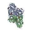
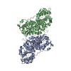
[English] 日本語
Yorodumi
- PDB-8hp2: CtPDC -

+
Open data


ID or keywords:

Loading...

-
Basic information

Entry
Database: PDB / ID: 8hp2
TitleCtPDC
ComponentsPyruvate decarboxylase
KeywordsLYASE / carboxylation / fold / unit
Function / homology
Function and homology information


carboxy-lyase activity / thiamine pyrophosphate binding / magnesium ion binding
Similarity search - Function
: / : / Thiamine pyrophosphate (TPP)-dependent enzyme / TPP-binding enzyme, conserved site / Thiamine pyrophosphate enzymes signature. / Thiamine pyrophosphate enzyme, central domain / Thiamine pyrophosphate enzyme, central domain / Thiamine pyrophosphate enzyme, N-terminal TPP-binding domain / Thiamine pyrophosphate enzyme, N-terminal TPP binding domain / Thiamine pyrophosphate enzyme, C-terminal TPP-binding ...: / : / Thiamine pyrophosphate (TPP)-dependent enzyme / TPP-binding enzyme, conserved site / Thiamine pyrophosphate enzymes signature. / Thiamine pyrophosphate enzyme, central domain / Thiamine pyrophosphate enzyme, central domain / Thiamine pyrophosphate enzyme, N-terminal TPP-binding domain / Thiamine pyrophosphate enzyme, N-terminal TPP binding domain / Thiamine pyrophosphate enzyme, C-terminal TPP-binding / Thiamine pyrophosphate enzyme, C-terminal TPP binding domain / Thiamin diphosphate-binding fold / DHS-like NAD/FAD-binding domain superfamily
Similarity search - Domain/homology
Pyruvate decarboxylase
Similarity search - Component
Biological speciesCandida tropicalis (yeast)
MethodX-RAY DIFFRACTION / MOLECULAR REPLACEMENT / Resolution: 3.05 Å
AuthorsXu, H.H. / Song, W.
Funding support1items
OrganizationGrant numberCountry
Not funded
CitationJournal: To Be Published
Title: Protein engineering of pyruvate decarboxylase to remove the rate-limiting bottleneck in the cascade pathway of tyrosol synthesis
Authors: Xu, H.H. / Song, W.
History
DepositionDec 11, 2022Deposition site: PDBJ / Processing site: PDBJ
Revision 1.0Jan 17, 2024Provider: repository / Type: Initial release

-
Structure visualization

Structure viewerMolecule:
MolmilJmol/JSmol

Downloads & links

-
Assembly

Deposited unit
A: Pyruvate decarboxylase
B: Pyruvate decarboxylase


Theoretical massNumber of molelcules
Total (without water)124,5092
Polymers124,5092
Non-polymers00
Water00
1
A: Pyruvate decarboxylase

B: Pyruvate decarboxylase


Theoretical massNumber of molelcules
Total (without water)124,5092
Polymers124,5092
Non-polymers00
Water0
TypeNameSymmetry operationNumber
identity operation1_555x,y,z1
crystal symmetry operation3_445x-1/2,y-1/2,z1
Buried area2530 Å2
ΔGint-14 kcal/mol
Surface area37220 Å2
MethodPISA
Unit cell
Length a, b, c (Å)123.471, 85.295, 113.534
Angle α, β, γ (deg.)90.00, 120.71, 90.00
Int Tables number5
Space group name H-MC121

-
Components

#1: Protein Pyruvate decarboxylase


Mass: 62254.344 Da / Num. of mol.: 2
Source method: isolated from a genetically manipulated source
Source: (gene. exp.) Candida tropicalis (yeast) / Gene: CTRG_03826 / Production host: Escherichia coli (E. coli) / References: UniProt: C5MDS4

-
Experimental details

-
Experiment

ExperimentMethod: X-RAY DIFFRACTION / Number of used crystals: 1

-
Sample preparation

CrystalDensity Matthews: 2.06 Å3/Da / Density % sol: 40.41 %
Crystal growTemperature: 293 K / Method: vapor diffusion, sitting drop
Details: 0.2 M sodium malonate pH 5.5, 18% Polyethylene glycol 3350

-
Data collection

DiffractionMean temperature: 100 K / Serial crystal experiment: N
Diffraction sourceSource: SEALED TUBE / Type: BRUKER D8 QUEST / Wavelength: 1.54178 Å
DetectorType: Bruker PHOTON II / Detector: PIXEL / Date: Jan 6, 2022
RadiationProtocol: SINGLE WAVELENGTH / Monochromatic (M) / Laue (L): M / Scattering type: x-ray
Radiation wavelengthWavelength: 1.54178 Å / Relative weight: 1
ReflectionResolution: 3.05→47.2 Å / Num. obs: 17253 / % possible obs: 89.3 % / Redundancy: 4 % / Rmerge(I) obs: 0.143 / Net I/σ(I): 8.9
Reflection shellResolution: 3.05→3.26 Å / Rmerge(I) obs: 0.558 / Num. unique obs: 3217

-
Processing

Software
NameClassification
Aimlessdata scaling
REFMACrefinement
RefinementMethod to determine structure: MOLECULAR REPLACEMENT
Starting model: 2VJY
Resolution: 3.05→47.2 Å / Cor.coef. Fo:Fc: 0.913 / Cor.coef. Fo:Fc free: 0.876 / SU B: 62.539 / SU ML: 0.487 / Cross valid method: THROUGHOUT / ESU R Free: 0.616 / Stereochemistry target values: MAXIMUM LIKELIHOOD / Details: HYDROGENS HAVE BEEN ADDED IN THE RIDING POSITIONS
RfactorNum. reflection% reflectionSelection details
Rfree0.28476 813 4.7 %RANDOM
Rwork0.2325 ---
obs0.23502 16438 88.53 %-
Solvent computationIon probe radii: 0.8 Å / Shrinkage radii: 0.8 Å / VDW probe radii: 1.2 Å / Solvent model: MASK
Displacement parametersBiso mean: 60.621 Å2
Baniso -1Baniso -2Baniso -3
1--1.91 Å20 Å20.81 Å2
2--1.34 Å2-0 Å2
3----0.23 Å2
Refinement stepCycle: 1 / Resolution: 3.05→47.2 Å
ProteinNucleic acidLigandSolventTotal
Num. atoms7392 0 0 0 7392
Refine LS restraints
Refine-IDTypeDev idealDev ideal targetNumber
X-RAY DIFFRACTIONr_bond_refined_d0.0030.0137540
X-RAY DIFFRACTIONr_bond_other_d0.0010.0177220
X-RAY DIFFRACTIONr_angle_refined_deg1.2261.63710241
X-RAY DIFFRACTIONr_angle_other_deg1.0591.57616642
X-RAY DIFFRACTIONr_dihedral_angle_1_deg6.025943
X-RAY DIFFRACTIONr_dihedral_angle_2_deg32.72923.788359
X-RAY DIFFRACTIONr_dihedral_angle_3_deg15.614151256
X-RAY DIFFRACTIONr_dihedral_angle_4_deg17.0261532
X-RAY DIFFRACTIONr_chiral_restr0.040.2996
X-RAY DIFFRACTIONr_gen_planes_refined0.0030.028516
X-RAY DIFFRACTIONr_gen_planes_other0.0010.021656
X-RAY DIFFRACTIONr_nbd_refined
X-RAY DIFFRACTIONr_nbd_other
X-RAY DIFFRACTIONr_nbtor_refined
X-RAY DIFFRACTIONr_nbtor_other
X-RAY DIFFRACTIONr_xyhbond_nbd_refined
X-RAY DIFFRACTIONr_xyhbond_nbd_other
X-RAY DIFFRACTIONr_metal_ion_refined
X-RAY DIFFRACTIONr_metal_ion_other
X-RAY DIFFRACTIONr_symmetry_vdw_refined
X-RAY DIFFRACTIONr_symmetry_vdw_other
X-RAY DIFFRACTIONr_symmetry_hbond_refined
X-RAY DIFFRACTIONr_symmetry_hbond_other
X-RAY DIFFRACTIONr_symmetry_metal_ion_refined
X-RAY DIFFRACTIONr_symmetry_metal_ion_other
X-RAY DIFFRACTIONr_mcbond_it0.8632.853811
X-RAY DIFFRACTIONr_mcbond_other0.8622.8493810
X-RAY DIFFRACTIONr_mcangle_it1.6284.264741
X-RAY DIFFRACTIONr_mcangle_other1.6284.2614742
X-RAY DIFFRACTIONr_scbond_it0.3562.833729
X-RAY DIFFRACTIONr_scbond_other0.3562.8313730
X-RAY DIFFRACTIONr_scangle_it
X-RAY DIFFRACTIONr_scangle_other0.7984.2475501
X-RAY DIFFRACTIONr_long_range_B_refined3.4233.2648244
X-RAY DIFFRACTIONr_long_range_B_other3.4233.2698245
X-RAY DIFFRACTIONr_rigid_bond_restr
X-RAY DIFFRACTIONr_sphericity_free
X-RAY DIFFRACTIONr_sphericity_bonded
Refine LS restraints NCS

Ens-ID: 1 / Number: 14811 / Refine-ID: X-RAY DIFFRACTION / Type: interatomic distance / Rms dev position: 0.06 Å / Weight position: 0.05

Dom-IDAuth asym-ID
1A
2B
LS refinement shellResolution: 3.05→3.129 Å / Total num. of bins used: 20
RfactorNum. reflection% reflection
Rfree0.329 75 -
Rwork0.308 1251 -
obs--91.76 %
Refinement TLS params.

Method: refined / Refine-ID: X-RAY DIFFRACTION

IDL112)L122)L132)L222)L232)L332)S11 (Å °)S12 (Å °)S13 (Å °)S21 (Å °)S22 (Å °)S23 (Å °)S31 (Å °)S32 (Å °)S33 (Å °)T112)T122)T132)T222)T232)T332)Origin x (Å)Origin y (Å)Origin z (Å)
13.0922-1.309-0.51741.50720.31632.46660.0163-0.02120.10450.1844-0.03450.11630.2478-0.28430.01820.3671-0.0309-0.02350.4725-0.10390.231-17.170212.904630.8175
21.6702-0.7005-0.40390.34280.07280.61550.0168-0.1005-0.00960.0523-0.07010.00080.013-0.04590.05330.5008-0.0349-0.00680.6093-0.04690.13682.44567.31445.3384
33.353-0.08091.05350.04610.15271.1621-0.01140.03270.06350.0774-0.0461-0.0030.31040.04830.05750.48190.03520.0070.48740.05970.08972.819-0.152318.2431
42.53350.3875-0.05124.0456-2.66681.8319-0.1515-0.3025-0.04290.33330.018-0.3412-0.18130.03280.13360.3968-0.0365-0.03050.5668-0.02990.216733.852224.6827.4428
51.38060.0466-0.13710.3969-0.080.0854-0.05010.3796-0.09690.2170.01290.1028-0.1617-0.04060.03730.42540.0547-0.02070.73810.0830.086910.689635.312718.8804
63.73920.6148-0.04840.3307-0.54831.393-0.16840.73640.3180.09760.15760.025-0.1149-0.27450.01080.4186-0.036-0.02650.6960.1040.056332.82837.95414.5764
Refinement TLS group
IDRefine-IDRefine TLS-IDAuth asym-IDAuth seq-ID
1X-RAY DIFFRACTION1A2 - 138
2X-RAY DIFFRACTION2A139 - 342
3X-RAY DIFFRACTION3A343 - 546
4X-RAY DIFFRACTION4B2 - 146
5X-RAY DIFFRACTION5B147 - 391
6X-RAY DIFFRACTION6B392 - 546

+
About Yorodumi

-
News

-
Feb 9, 2022. New format data for meta-information of EMDB entries

New format data for meta-information of EMDB entries

  • Version 3 of the EMDB header file is now the official format.
  • The previous official version 1.9 will be removed from the archive.

Related info.:EMDB header

External links:wwPDB to switch to version 3 of the EMDB data model

-
Aug 12, 2020. Covid-19 info

Covid-19 info

URL: https://pdbjlvh1.pdbj.org/emnavi/covid19.php

New page: Covid-19 featured information page in EM Navigator.

Related info.:Covid-19 info / Mar 5, 2020. Novel coronavirus structure data

+
Mar 5, 2020. Novel coronavirus structure data

Novel coronavirus structure data

Related info.:Yorodumi Speices / Aug 12, 2020. Covid-19 info

External links:COVID-19 featured content - PDBj / Molecule of the Month (242):Coronavirus Proteases

+
Jan 31, 2019. EMDB accession codes are about to change! (news from PDBe EMDB page)

EMDB accession codes are about to change! (news from PDBe EMDB page)

  • The allocation of 4 digits for EMDB accession codes will soon come to an end. Whilst these codes will remain in use, new EMDB accession codes will include an additional digit and will expand incrementally as the available range of codes is exhausted. The current 4-digit format prefixed with “EMD-” (i.e. EMD-XXXX) will advance to a 5-digit format (i.e. EMD-XXXXX), and so on. It is currently estimated that the 4-digit codes will be depleted around Spring 2019, at which point the 5-digit format will come into force.
  • The EM Navigator/Yorodumi systems omit the EMD- prefix.

Related info.:Q: What is EMD? / ID/Accession-code notation in Yorodumi/EM Navigator

External links:EMDB Accession Codes are Changing Soon! / Contact to PDBj

+
Jul 12, 2017. Major update of PDB

Major update of PDB

  • wwPDB released updated PDB data conforming to the new PDBx/mmCIF dictionary.
  • This is a major update changing the version number from 4 to 5, and with Remediation, in which all the entries are updated.
  • In this update, many items about electron microscopy experimental information are reorganized (e.g. em_software).
  • Now, EM Navigator and Yorodumi are based on the updated data.

External links:wwPDB Remediation / Enriched Model Files Conforming to OneDep Data Standards Now Available in the PDB FTP Archive

-
Yorodumi

Thousand views of thousand structures

  • Yorodumi is a browser for structure data from EMDB, PDB, SASBDB, etc.
  • This page is also the successor to EM Navigator detail page, and also detail information page/front-end page for Omokage search.
  • The word "yorodu" (or yorozu) is an old Japanese word meaning "ten thousand". "mi" (miru) is to see.

Related info.:EMDB / PDB / SASBDB / Comparison of 3 databanks / Yorodumi Search / Aug 31, 2016. New EM Navigator & Yorodumi / Yorodumi Papers / Jmol/JSmol / Function and homology information / Changes in new EM Navigator and Yorodumi

Read more