 Movie
Movie Controller


+ Open data

- Basic information
| Entry | Database: PDB / ID: 8hoe | ||||||
|---|---|---|---|---|---|---|---|
| Title | Apo structure of HopBF1 kinase from Ewingella americana | ||||||
 Components Components | Type III effector HopBF1 | ||||||
 Keywords Keywords | TRANSFERASE / Kinase | ||||||
| Function / homology | : / Transferases; Transferring phosphorus-containing groups; Phosphotransferases with an alcohol group as acceptor / kinase activity / host cell / extracellular region / ATP binding / Type III effector protein HopBF1 Function and homology information Function and homology information | ||||||
| Biological species |  Ewingella americana (bacteria) Ewingella americana (bacteria) | ||||||
| Method |  X-RAY DIFFRACTION / SYNCHROTRON / MOLECULAR REPLACEMENT / Resolution: 2.2 Å X-RAY DIFFRACTION / SYNCHROTRON / MOLECULAR REPLACEMENT / Resolution: 2.2 Å | ||||||
 Authors Authors | Wang, C.C. / Xu, T. | ||||||
| Funding support |  China, 1items China, 1items
| ||||||
 Citation Citation | Journal: To Be Published Title: Apo structure of HopBF1 kinase from Ewingella americana Authors: Wang, C.C. | ||||||
| History |
|
- Structure visualization
| Structure viewer | Molecule: MolmilJmol/JSmol |
|---|
- Downloads & links
-Download
| PDBx/mmCIF format | 8hoe.cif.gz | 89.8 KB | Display | PDBx/mmCIF format |
|---|---|---|---|---|
| PDB format | pdb8hoe.ent.gz | 64.7 KB | Display | PDB format |
| PDBx/mmJSON format | 8hoe.json.gz | Tree view | PDBx/mmJSON format | |
| Others |  Other downloads Other downloads |
-Validation report
| Arichive directory | https://data.pdbj.org/pub/pdb/validation_reports/ho/8hoeftp://data.pdbj.org/pub/pdb/validation_reports/ho/8hoe | HTTPS FTP |
|---|
-Related structure data
| Similar structure data |
|---|
-Links
 PDBj
PDBj- Assembly
| Deposited unit | 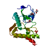
| ||||||||||
|---|---|---|---|---|---|---|---|---|---|---|---|
| 1 |
| ||||||||||
| Unit cell |
|
-Components
| #1: Protein | Mass: 21166.791 Da / Num. of mol.: 1 Source method: isolated from a genetically manipulated source Source: (gene. exp.) Ewingella americana (bacteria) / Gene: GEAM_1328 / Production host: | ||||||
|---|---|---|---|---|---|---|---|
| #2: Chemical | ChemComp-EDO /   Mass: 62.068 Da / Num. of mol.: 5 / Source method: isolated from a natural source / Formula: C2H6O2 Mass: 62.068 Da / Num. of mol.: 5 / Source method: isolated from a natural source / Formula: C2H6O2#3: Chemical | ChemComp-SO4 / |   Mass: 96.063 Da / Num. of mol.: 1 / Source method: obtained synthetically / Formula: SO4 Mass: 96.063 Da / Num. of mol.: 1 / Source method: obtained synthetically / Formula: SO4#4: Water | ChemComp-HOH / |  Mass: 18.015 Da / Num. of mol.: 31 / Source method: isolated from a natural source / Formula: H2O Mass: 18.015 Da / Num. of mol.: 31 / Source method: isolated from a natural source / Formula: H2OHas ligand of interest | N | |
-Experimental details
-Experiment
| Experiment | Method: X-RAY DIFFRACTION / Number of used crystals: 1 |
|---|
- Sample preparation
| Crystal | Density Matthews: 5.08 Å3/Da / Density % sol: 75.78 % |
|---|---|
| Crystal grow | Temperature: 289.15 K / Method: vapor diffusion, sitting drop Details: 2 M ammonium sulfate, 8 % (V/V) ethylene glycol, ,10 % (w/v) PEG 8000, 0.1 M HEPES sodium pH 7.5 |
-Data collection
| Diffraction | Mean temperature: 100 K / Serial crystal experiment: N |
|---|---|
| Diffraction source | Source: SYNCHROTRON / Site: SSRF / Beamline: BL19U1 / Wavelength: 0.97852 Å |
| Detector | Type: DECTRIS PILATUS 6M / Detector: PIXEL / Date: May 29, 2020 |
| Radiation | Protocol: SINGLE WAVELENGTH / Monochromatic (M) / Laue (L): M / Scattering type: x-ray |
| Radiation wavelength | Wavelength: 0.97852 Å / Relative weight: 1 |
| Reflection | Resolution: 2.2→38.76 Å / Num. obs: 21950 / % possible obs: 99.96 % / Redundancy: 19.2 % / Biso Wilson estimate: 58.63 Å2 / CC1/2: 1 / CC star: 1 / Rmerge(I) obs: 0.067 / Rpim(I) all: 0.016 / Rrim(I) all: 0.069 / Net I/σ(I): 23.88 |
| Reflection shell | Resolution: 2.2→2.3 Å / Rmerge(I) obs: 1.441 / Num. unique obs: 2681 / CC1/2: 0.755 / CC star: 0.928 / Rpim(I) all: 0.348 |
- Processing
| Software |
| |||||||||||||||||||||||||||||||||||||||||||||||||||||||||||||||
|---|---|---|---|---|---|---|---|---|---|---|---|---|---|---|---|---|---|---|---|---|---|---|---|---|---|---|---|---|---|---|---|---|---|---|---|---|---|---|---|---|---|---|---|---|---|---|---|---|---|---|---|---|---|---|---|---|---|---|---|---|---|---|---|---|
| Refinement | Method to determine structure: MOLECULAR REPLACEMENT / Resolution: 2.2→38.76 Å / SU ML: 0.3332 / Cross valid method: FREE R-VALUE / σ(F): 1.36 / Phase error: 25.1306 Stereochemistry target values: GeoStd + Monomer Library + CDL v1.2
| |||||||||||||||||||||||||||||||||||||||||||||||||||||||||||||||
| Solvent computation | Shrinkage radii: 0.9 Å / VDW probe radii: 1.1 Å / Solvent model: FLAT BULK SOLVENT MODEL | |||||||||||||||||||||||||||||||||||||||||||||||||||||||||||||||
| Displacement parameters | Biso mean: 70.34 Å2 | |||||||||||||||||||||||||||||||||||||||||||||||||||||||||||||||
| Refinement step | Cycle: LAST / Resolution: 2.2→38.76 Å
| |||||||||||||||||||||||||||||||||||||||||||||||||||||||||||||||
| Refine LS restraints |
| |||||||||||||||||||||||||||||||||||||||||||||||||||||||||||||||
| LS refinement shell |
|
