 Movie
Movie Controller


+ Open data

- Basic information
| Entry | Database: PDB / ID: 8hly | ||||||
|---|---|---|---|---|---|---|---|
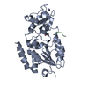
| Title | Crystal structure of SIRT3 in complex with H3K23la peptide | ||||||
 Components Components |
| ||||||
 Keywords Keywords | HYDROLASE / SIRT3 / Lysine lactylation eraser | ||||||
| Function / homology |  Function and homology information Function and homology informationpositive regulation of catalase activity / positive regulation of superoxide dismutase activity / NAD-dependent protein lysine delactylase activity / positive regulation of ceramide biosynthetic process / peptidyl-lysine deacetylation / Maturation of TCA enzymes and regulation of TCA cycle / protein acetyllysine N-acetyltransferase / NAD-dependent protein lysine deacetylase activity / protein deacetylation / histone deacetylase activity, NAD-dependent ...positive regulation of catalase activity / positive regulation of superoxide dismutase activity / NAD-dependent protein lysine delactylase activity / positive regulation of ceramide biosynthetic process / peptidyl-lysine deacetylation / Maturation of TCA enzymes and regulation of TCA cycle / protein acetyllysine N-acetyltransferase / NAD-dependent protein lysine deacetylase activity / protein deacetylation / histone deacetylase activity, NAD-dependent / positive regulation of oxidative phosphorylation / Regulation of FOXO transcriptional activity by acetylation / protein lysine deacetylase activity / cellular response to stress / negative regulation of reactive oxygen species metabolic process / NAD+ binding / FOXO-mediated transcription of oxidative stress, metabolic and neuronal genes / Mitochondrial unfolded protein response (UPRmt) / Transferases; Acyltransferases; Transferring groups other than aminoacyl groups / aerobic respiration / Transcriptional activation of mitochondrial biogenesis / negative regulation of ERK1 and ERK2 cascade / positive regulation of insulin secretion / sequence-specific DNA binding / mitochondrial matrix / enzyme binding / protein-containing complex / mitochondrion / zinc ion binding / nucleoplasm / nucleus Similarity search - Function | ||||||
| Biological species |  Homo sapiens (human) Homo sapiens (human) | ||||||
| Method |  X-RAY DIFFRACTION / SYNCHROTRON / MOLECULAR REPLACEMENT / Resolution: 2 Å X-RAY DIFFRACTION / SYNCHROTRON / MOLECULAR REPLACEMENT / Resolution: 2 Å | ||||||
 Authors Authors | Zhuming, F. / Hao, Q. | ||||||
| Funding support |  Hong Kong, 1items Hong Kong, 1items
| ||||||
 Citation Citation | Journal: Iscience / Year: 2023 Title: Identification of SIRT3 as an eraser of H4K16la. Authors: Fan, Z. / Liu, Z. / Zhang, N. / Wei, W. / Cheng, K. / Sun, H. / Hao, Q. | ||||||
| History |
|
- Structure visualization
| Structure viewer | Molecule: MolmilJmol/JSmol |
|---|
- Downloads & links
-Download
| PDBx/mmCIF format | 8hly.cif.gz | 180.4 KB | Display | PDBx/mmCIF format |
|---|---|---|---|---|
| PDB format | pdb8hly.ent.gz | 143.2 KB | Display | PDB format |
| PDBx/mmJSON format | 8hly.json.gz | Tree view | PDBx/mmJSON format | |
| Others |  Other downloads Other downloads |
-Validation report
| Arichive directory | https://data.pdbj.org/pub/pdb/validation_reports/hl/8hlyftp://data.pdbj.org/pub/pdb/validation_reports/hl/8hly | HTTPS FTP |
|---|
-Related structure data
-Links
 PDBj
PDBj


- Assembly
| Deposited unit | 
| ||||||||
|---|---|---|---|---|---|---|---|---|---|
| 1 |
| ||||||||
| Unit cell |
|
-Components
-Protein / Protein/peptide , 2 types, 2 molecules AB
| #1: Protein | Mass: 31253.010 Da / Num. of mol.: 1 Source method: isolated from a genetically manipulated source Source: (gene. exp.) Homo sapiens (human) / Gene: SIRT3, SIR2L3 / Production host:  References: UniProt: Q9NTG7, protein acetyllysine N-acetyltransferase |
|---|---|
| #2: Protein/peptide | Mass: 618.726 Da / Num. of mol.: 1 / Source method: obtained synthetically Details: residues 21-26 of histone H3, with lactylated Lys-23 Source: (synth.) Homo sapiens (human) |
-Non-polymers , 5 types, 175 molecules 


| #3: Chemical |  Mass: 92.094 Da / Num. of mol.: 3 / Source method: obtained synthetically / Formula: C3H8O3 Mass: 92.094 Da / Num. of mol.: 3 / Source method: obtained synthetically / Formula: C3H8O3#4: Chemical | ChemComp-EDO / |  Mass: 62.068 Da / Num. of mol.: 1 / Source method: obtained synthetically / Formula: C2H6O2 Mass: 62.068 Da / Num. of mol.: 1 / Source method: obtained synthetically / Formula: C2H6O2#5: Chemical | ChemComp-ZN / |  Mass: 65.409 Da / Num. of mol.: 1 / Source method: isolated from a natural source / Formula: Zn Mass: 65.409 Da / Num. of mol.: 1 / Source method: isolated from a natural source / Formula: Zn#6: Chemical | ChemComp-2OP / ( |  Mass: 90.078 Da / Num. of mol.: 1 / Source method: obtained synthetically / Formula: C3H6O3 / Feature type: SUBJECT OF INVESTIGATION Mass: 90.078 Da / Num. of mol.: 1 / Source method: obtained synthetically / Formula: C3H6O3 / Feature type: SUBJECT OF INVESTIGATION#7: Water | ChemComp-HOH / | Mass: 18.015 Da / Num. of mol.: 169 / Source method: isolated from a natural source / Formula: H2O |
|---|
-Details
| Has ligand of interest | Y |
|---|
-Experimental details
-Experiment
| Experiment | Method: X-RAY DIFFRACTION / Number of used crystals: 1 |
|---|
- Sample preparation
| Crystal | Density Matthews: 2 Å3/Da / Density % sol: 38.44 % |
|---|---|
| Crystal grow | Temperature: 291 K / Method: vapor diffusion, sitting drop / pH: 6.5 Details: 0.1 M BIS-TRIS pH at 6.5, 28% (w/v) Polyethylene glycol monomethyl ether 2000 |
-Data collection
| Diffraction | Mean temperature: 100 K / Serial crystal experiment: N |
|---|---|
| Diffraction source | Source: SYNCHROTRON / Site: SSRF / Beamline: BL19U1 / Wavelength: 0.97847 Å |
| Detector | Type: DECTRIS PILATUS 6M / Detector: PIXEL / Date: Feb 1, 2020 |
| Radiation | Protocol: SINGLE WAVELENGTH / Monochromatic (M) / Laue (L): M / Scattering type: x-ray |
| Radiation wavelength | Wavelength: 0.97847 Å / Relative weight: 1 |
| Reflection | Resolution: 1.998→44.593 Å / Num. obs: 25433 / % possible obs: 95.93 % / Redundancy: 1.5 % / Biso Wilson estimate: 22.99 Å2 / CC1/2: 0.996 / CC star: 0.999 / Rmerge(I) obs: 0.03378 / Rpim(I) all: 0.03378 / Rrim(I) all: 0.04777 / Net I/σ(I): 15.12 |
| Reflection shell | Resolution: 1.998→2.07 Å / Redundancy: 1 % / Rmerge(I) obs: 0.07989 / Mean I/σ(I) obs: 3.48 / Num. unique obs: 1294 / CC1/2: 1 / CC star: 1 / Rpim(I) all: 0.07989 / Rrim(I) all: 0.113 / % possible all: 72.33 |
- Processing
| Software |
| |||||||||||||||||||||||||||||||||||||||||||||||||||||||||||||||||||||||||||||||||||||||||||||||||||||||||||||||||||||||||||||||||||||
|---|---|---|---|---|---|---|---|---|---|---|---|---|---|---|---|---|---|---|---|---|---|---|---|---|---|---|---|---|---|---|---|---|---|---|---|---|---|---|---|---|---|---|---|---|---|---|---|---|---|---|---|---|---|---|---|---|---|---|---|---|---|---|---|---|---|---|---|---|---|---|---|---|---|---|---|---|---|---|---|---|---|---|---|---|---|---|---|---|---|---|---|---|---|---|---|---|---|---|---|---|---|---|---|---|---|---|---|---|---|---|---|---|---|---|---|---|---|---|---|---|---|---|---|---|---|---|---|---|---|---|---|---|---|---|
| Refinement | Method to determine structure: MOLECULAR REPLACEMENT / Resolution: 2→44.59 Å / SU ML: 0.18 / Cross valid method: FREE R-VALUE / σ(F): 1.34 / Phase error: 19.2 / Stereochemistry target values: ML
| |||||||||||||||||||||||||||||||||||||||||||||||||||||||||||||||||||||||||||||||||||||||||||||||||||||||||||||||||||||||||||||||||||||
| Solvent computation | Shrinkage radii: 0.9 Å / VDW probe radii: 1.11 Å / Solvent model: FLAT BULK SOLVENT MODEL | |||||||||||||||||||||||||||||||||||||||||||||||||||||||||||||||||||||||||||||||||||||||||||||||||||||||||||||||||||||||||||||||||||||
| Refinement step | Cycle: LAST / Resolution: 2→44.59 Å
| |||||||||||||||||||||||||||||||||||||||||||||||||||||||||||||||||||||||||||||||||||||||||||||||||||||||||||||||||||||||||||||||||||||
| Refine LS restraints |
| |||||||||||||||||||||||||||||||||||||||||||||||||||||||||||||||||||||||||||||||||||||||||||||||||||||||||||||||||||||||||||||||||||||
| LS refinement shell |
| |||||||||||||||||||||||||||||||||||||||||||||||||||||||||||||||||||||||||||||||||||||||||||||||||||||||||||||||||||||||||||||||||||||
| Refinement TLS params. | Method: refined / Origin x: -4.8497 Å / Origin y: -18.3698 Å / Origin z: 19.6547 Å
| |||||||||||||||||||||||||||||||||||||||||||||||||||||||||||||||||||||||||||||||||||||||||||||||||||||||||||||||||||||||||||||||||||||
| Refinement TLS group | Selection details: all |
