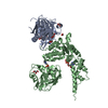
 Movie
Movie Controller

[English] 日本語
 Yorodumi
- PDB-8hl6: Crystal structure of human valosin-containing protein methyltrans... -
+ Open data

- Basic information
| Entry | Database: PDB / ID: 8hl6 | |||||||||
|---|---|---|---|---|---|---|---|---|---|---|
| Title | Crystal structure of human valosin-containing protein methyltransferase | |||||||||
 Components Components | Protein N-lysine methyltransferase METTL21D | |||||||||
 Keywords Keywords | PROTEIN BINDING / VCPKMT / methyltransferase / ASPL / ASPSCR1 / UBXD9 / TUG | |||||||||
| Function / homology |  Function and homology information Function and homology informationpeptidyl-lysine methylation / peptidyl-lysine trimethylation / negative regulation of ATP-dependent activity / protein-lysine N-methyltransferase activity / Protein methylation / Transferases; Transferring one-carbon groups; Methyltransferases / ATPase binding / protein-containing complex / cytoplasm / cytosol Similarity search - Function | |||||||||
| Biological species |  Homo sapiens (human) Homo sapiens (human) | |||||||||
| Method |  X-RAY DIFFRACTION / SYNCHROTRON / MOLECULAR REPLACEMENT / Resolution: 1.8 Å X-RAY DIFFRACTION / SYNCHROTRON / MOLECULAR REPLACEMENT / Resolution: 1.8 Å | |||||||||
 Authors Authors | Kang, W. / Yang, J.K. | |||||||||
| Funding support |  Korea, Republic Of, 2items Korea, Republic Of, 2items
| |||||||||
 Citation Citation | Journal: Iscience / Year: 2023 Title: Structural basis for recognition and methylation of p97 by METTL21D, a valosin-containing protein lysine methyltransferase. Authors: Nguyen, T.Q. / Koh, S. / Kwon, J. / Jang, S. / Kang, W. / Yang, J.K. | |||||||||
| History |
|
- Structure visualization
| Structure viewer | Molecule: MolmilJmol/JSmol |
|---|
- Downloads & links
-Download
| PDBx/mmCIF format | 8hl6.cif.gz | 104.7 KB | Display | PDBx/mmCIF format |
|---|---|---|---|---|
| PDB format | pdb8hl6.ent.gz | 77.6 KB | Display | PDB format |
| PDBx/mmJSON format | 8hl6.json.gz | Tree view | PDBx/mmJSON format | |
| Others |  Other downloads Other downloads |
-Validation report
| Summary document | 8hl6_validation.pdf.gz | 708.2 KB | Display | wwPDB validaton report |
|---|---|---|---|---|
| Full document | 8hl6_full_validation.pdf.gz | 708.9 KB | Display | |
| Data in XML | 8hl6_validation.xml.gz | 10.9 KB | Display | |
| Data in CIF | 8hl6_validation.cif.gz | 14.8 KB | Display | |
| Arichive directory | https://data.pdbj.org/pub/pdb/validation_reports/hl/8hl6ftp://data.pdbj.org/pub/pdb/validation_reports/hl/8hl6 | HTTPS FTP |
-Related structure data
-Links
 PDBj
PDBj


- Assembly
| Deposited unit | 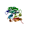
| ||||||||
|---|---|---|---|---|---|---|---|---|---|
| 1 |
| ||||||||
| Unit cell |
|
-Components
| #1: Protein | Mass: 26665.486 Da / Num. of mol.: 1 Source method: isolated from a genetically manipulated source Source: (gene. exp.) Homo sapiens (human) / Gene: METTL21D / Production host:  References: UniProt: Q9H867, Transferases; Transferring one-carbon groups; Methyltransferases |
|---|---|
| #2: Chemical | ChemComp-SAM /   Mass: 398.437 Da / Num. of mol.: 1 / Source method: obtained synthetically / Formula: C15H22N6O5S / Feature type: SUBJECT OF INVESTIGATION Mass: 398.437 Da / Num. of mol.: 1 / Source method: obtained synthetically / Formula: C15H22N6O5S / Feature type: SUBJECT OF INVESTIGATION |
| #3: Water | ChemComp-HOH /  Mass: 18.015 Da / Num. of mol.: 96 / Source method: isolated from a natural source / Formula: H2O Mass: 18.015 Da / Num. of mol.: 96 / Source method: isolated from a natural source / Formula: H2O |
| Has ligand of interest | Y |
-Experimental details
-Experiment
| Experiment | Method: X-RAY DIFFRACTION / Number of used crystals: 1 |
|---|
- Sample preparation
| Crystal | Density Matthews: 2.44 Å3/Da / Density % sol: 49.67 % |
|---|---|
| Crystal grow | Temperature: 295 K / Method: vapor diffusion, hanging drop Details: 20 % PEG 1000, 0.1 M Na/K phosphate (pH 6.2), and 0.2M sodium chloride PH range: 6.2 |
-Data collection
| Diffraction | Mean temperature: 100 K / Serial crystal experiment: N |
|---|---|
| Diffraction source | Source: SYNCHROTRON / Site: PAL/PLS / Beamline: 5C (4A) / Wavelength: 1 Å |
| Detector | Type: ADSC QUANTUM 315r / Detector: CCD / Date: Jan 10, 2013 |
| Radiation | Protocol: SINGLE WAVELENGTH / Monochromatic (M) / Laue (L): M / Scattering type: x-ray |
| Radiation wavelength | Wavelength: 1 Å / Relative weight: 1 |
| Reflection | Resolution: 1.8→64.6 Å / Num. obs: 23428 / % possible obs: 93.2 % / Redundancy: 9.4 % / CC1/2: 0.999 / Rpim(I) all: 0.023 / Net I/σ(I): 17.2 |
| Reflection shell | Resolution: 1.8→1.9 Å / Num. unique obs: 2011 / CC1/2: 0.405 / Rpim(I) all: 0.683 |
- Processing
| Software |
| |||||||||||||||||||||||||||||||||||||||||||||||||||||||||||||||
|---|---|---|---|---|---|---|---|---|---|---|---|---|---|---|---|---|---|---|---|---|---|---|---|---|---|---|---|---|---|---|---|---|---|---|---|---|---|---|---|---|---|---|---|---|---|---|---|---|---|---|---|---|---|---|---|---|---|---|---|---|---|---|---|---|
| Refinement | Method to determine structure: MOLECULAR REPLACEMENT / Resolution: 1.8→32.748 Å / SU ML: 0.25 / Cross valid method: FREE R-VALUE / σ(F): 1.34 / Phase error: 26.5 / Stereochemistry target values: ML
| |||||||||||||||||||||||||||||||||||||||||||||||||||||||||||||||
| Solvent computation | Shrinkage radii: 0.9 Å / VDW probe radii: 1.11 Å / Solvent model: FLAT BULK SOLVENT MODEL | |||||||||||||||||||||||||||||||||||||||||||||||||||||||||||||||
| Refinement step | Cycle: LAST / Resolution: 1.8→32.748 Å
| |||||||||||||||||||||||||||||||||||||||||||||||||||||||||||||||
| Refine LS restraints |
| |||||||||||||||||||||||||||||||||||||||||||||||||||||||||||||||
| LS refinement shell |
| |||||||||||||||||||||||||||||||||||||||||||||||||||||||||||||||
| Refinement TLS params. | Method: refined / Origin x: -25.701 Å / Origin y: 6.4623 Å / Origin z: 8.3012 Å
| |||||||||||||||||||||||||||||||||||||||||||||||||||||||||||||||
| Refinement TLS group | Selection details: all |
