 Movie
Movie Controller


+ Open data

- Basic information
| Entry | Database: PDB / ID: 8hgr | ||||||
|---|---|---|---|---|---|---|---|
| Title | The apo-flavodoxin monomer from Synechococcus elongatus PCC 7942 | ||||||
 Components Components | Flavodoxin | ||||||
 Keywords Keywords | ELECTRON TRANSPORT / FLAVODOXIN / phosphate BINDING / REDOX POTENTIAL / FMN BINDING | ||||||
| Function / homology |  Function and homology information Function and homology information | ||||||
| Biological species |  Synechococcus elongatus PCC 7942 = FACHB-805 (bacteria) Synechococcus elongatus PCC 7942 = FACHB-805 (bacteria) | ||||||
| Method |  X-RAY DIFFRACTION / SYNCHROTRON / MOLECULAR REPLACEMENT / Resolution: 1.84 Å X-RAY DIFFRACTION / SYNCHROTRON / MOLECULAR REPLACEMENT / Resolution: 1.84 Å | ||||||
 Authors Authors | Liu, S.W. / Chen, Y.Y. / Gong, Y. / Cao, P. | ||||||
| Funding support |  China, 1items China, 1items
| ||||||
 Citation Citation | Journal: Biochem.Biophys.Res.Commun. / Year: 2022 Title: A dimer-monomer transition captured by the crystal structures of cyanobacterial apo flavodoxin. Authors: Liu, S. / Chen, Y. / Du, T. / Zhao, W. / Liu, X. / Zhang, H. / Yuan, Q. / Gao, L. / Dong, Y. / Gao, X. / Gong, Y. / Cao, P. | ||||||
| History |
|
- Structure visualization
| Structure viewer | Molecule: MolmilJmol/JSmol |
|---|
- Downloads & links
-Download
| PDBx/mmCIF format | 8hgr.cif.gz | 92 KB | Display | PDBx/mmCIF format |
|---|---|---|---|---|
| PDB format | pdb8hgr.ent.gz | 63.3 KB | Display | PDB format |
| PDBx/mmJSON format | 8hgr.json.gz | Tree view | PDBx/mmJSON format | |
| Others |  Other downloads Other downloads |
-Validation report
| Summary document | 8hgr_validation.pdf.gz | 427.8 KB | Display | wwPDB validaton report |
|---|---|---|---|---|
| Full document | 8hgr_full_validation.pdf.gz | 427.9 KB | Display | |
| Data in XML | 8hgr_validation.xml.gz | 9.7 KB | Display | |
| Data in CIF | 8hgr_validation.cif.gz | 13.2 KB | Display | |
| Arichive directory | https://data.pdbj.org/pub/pdb/validation_reports/hg/8hgrftp://data.pdbj.org/pub/pdb/validation_reports/hg/8hgr | HTTPS FTP |
-Related structure data
| Related structure data |  8hgqC 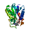 1cznS S: Starting model for refinement C: citing same article ( |
|---|---|
| Similar structure data |
-Links
 PDBj
PDBj

- Assembly
| Deposited unit | 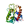
| ||||||||||||
|---|---|---|---|---|---|---|---|---|---|---|---|---|---|
| 1 |
| ||||||||||||
| Unit cell |
|
-Components
| #1: Protein | Mass: 20781.623 Da / Num. of mol.: 1 Source method: isolated from a genetically manipulated source Source: (gene. exp.) Synechococcus elongatus PCC 7942 = FACHB-805 (bacteria)Strain: PCC 7942 / FACHB-805 / Gene: isiB, Synpcc7942_1541 / Production host: |
|---|---|
| #2: Chemical | ChemComp-MG /   Mass: 24.305 Da / Num. of mol.: 1 / Source method: obtained synthetically / Formula: Mg Mass: 24.305 Da / Num. of mol.: 1 / Source method: obtained synthetically / Formula: Mg |
| #3: Chemical | ChemComp-CL /   Mass: 35.453 Da / Num. of mol.: 1 / Source method: isolated from a natural source / Formula: Cl Mass: 35.453 Da / Num. of mol.: 1 / Source method: isolated from a natural source / Formula: Cl |
| #4: Water | ChemComp-HOH /  Mass: 18.015 Da / Num. of mol.: 136 / Source method: isolated from a natural source / Formula: H2O Mass: 18.015 Da / Num. of mol.: 136 / Source method: isolated from a natural source / Formula: H2O |
| Has ligand of interest | N |
-Experimental details
-Experiment
| Experiment | Method: X-RAY DIFFRACTION / Number of used crystals: 1 |
|---|
- Sample preparation
| Crystal | Density Matthews: 2.12 Å3/Da / Density % sol: 41.97 % |
|---|---|
| Crystal grow | Temperature: 277 K / Method: vapor diffusion, sitting drop / pH: 8.5 Details: 0.2 M Magnesium chloride; 0.1 M Tris-HCl pH8.5; 30% PEG4000 |
-Data collection
| Diffraction | Mean temperature: 100 K / Serial crystal experiment: N |
|---|---|
| Diffraction source | Source: SYNCHROTRON / Site: SSRF / Beamline: BL18U1 / Wavelength: 0.98 Å |
| Detector | Type: DECTRIS PILATUS3 S 6M / Detector: PIXEL / Date: Feb 14, 2022 |
| Radiation | Protocol: SINGLE WAVELENGTH / Monochromatic (M) / Laue (L): M / Scattering type: x-ray |
| Radiation wavelength | Wavelength: 0.98 Å / Relative weight: 1 |
| Reflection | Resolution: 1.84→50 Å / Num. obs: 15654 / % possible obs: 97.5 % / Redundancy: 11.6 % / Biso Wilson estimate: 23.79 Å2 / Rmerge(I) obs: 0.095 / Rpim(I) all: 0.03 / Net I/σ(I): 15.6 |
| Reflection shell | Resolution: 1.84→1.93 Å / Rmerge(I) obs: 0.675 / Mean I/σ(I) obs: 2.3 / Num. unique obs: 1906 / Rpim(I) all: 0.298 / % possible all: 83.1 |
- Processing
| Software |
| ||||||||||||||||||||||||||||||||||||||||||||||||||||||||||||||||||||||||||||||||||||
|---|---|---|---|---|---|---|---|---|---|---|---|---|---|---|---|---|---|---|---|---|---|---|---|---|---|---|---|---|---|---|---|---|---|---|---|---|---|---|---|---|---|---|---|---|---|---|---|---|---|---|---|---|---|---|---|---|---|---|---|---|---|---|---|---|---|---|---|---|---|---|---|---|---|---|---|---|---|---|---|---|---|---|---|---|---|
| Refinement | Method to determine structure: MOLECULAR REPLACEMENT Starting model: 1CZN Resolution: 1.84→40.84 Å / SU ML: 0.1726 / Cross valid method: FREE R-VALUE / σ(F): 100 / Phase error: 19.2415 Stereochemistry target values: GeoStd + Monomer Library + CDL v1.2
| ||||||||||||||||||||||||||||||||||||||||||||||||||||||||||||||||||||||||||||||||||||
| Solvent computation | Shrinkage radii: 0.9 Å / VDW probe radii: 1.11 Å / Solvent model: FLAT BULK SOLVENT MODEL | ||||||||||||||||||||||||||||||||||||||||||||||||||||||||||||||||||||||||||||||||||||
| Displacement parameters | Biso mean: 28.09 Å2 | ||||||||||||||||||||||||||||||||||||||||||||||||||||||||||||||||||||||||||||||||||||
| Refinement step | Cycle: LAST / Resolution: 1.84→40.84 Å
| ||||||||||||||||||||||||||||||||||||||||||||||||||||||||||||||||||||||||||||||||||||
| Refine LS restraints |
| ||||||||||||||||||||||||||||||||||||||||||||||||||||||||||||||||||||||||||||||||||||
| LS refinement shell |
|
