 Movie
Movie Controller


+ Open data

- Basic information
| Entry | Database: PDB / ID: 8hfm | |||||||||
|---|---|---|---|---|---|---|---|---|---|---|
| Title | Crystal Structure of Mycobacterium smegmatis MshC | |||||||||
 Components Components | L-cysteine:1D-myo-inositol 2-amino-2-deoxy-alpha-D-glucopyranoside ligase | |||||||||
 Keywords Keywords | LIGASE / Rossmann fold | |||||||||
| Function / homology |  Function and homology information Function and homology informationL-cysteine:1D-myo-inositol 2-amino-2-deoxy-alpha-D-glucopyranoside ligase / cysteine-glucosaminylinositol ligase activity / cysteine-tRNA ligase activity / cysteinyl-tRNA aminoacylation / mycothiol biosynthetic process / zinc ion binding / ATP binding / cytosol Similarity search - Function | |||||||||
| Biological species |  Mycolicibacterium smegmatis (bacteria) Mycolicibacterium smegmatis (bacteria) | |||||||||
| Method |  X-RAY DIFFRACTION / SYNCHROTRON / MOLECULAR REPLACEMENT / Resolution: 2.41 Å X-RAY DIFFRACTION / SYNCHROTRON / MOLECULAR REPLACEMENT / Resolution: 2.41 Å | |||||||||
 Authors Authors | Pang, L. / Weeks, S.D. / Strelkov, S.V. | |||||||||
| Funding support |  Belgium, 2items Belgium, 2items
| |||||||||
 Citation Citation | Journal: Int J Mol Sci / Year: 2022 Title: Structural Basis of Cysteine Ligase MshC Inhibition by Cysteinyl-Sulfonamides. Authors: Pang, L. / Lenders, S. / Osipov, E.M. / Weeks, S.D. / Rozenski, J. / Piller, T. / Cappoen, D. / Strelkov, S.V. / Van Aerschot, A. | |||||||||
| History |
|
- Structure visualization
| Structure viewer | Molecule: MolmilJmol/JSmol |
|---|
- Downloads & links
-Download
| PDBx/mmCIF format | 8hfm.cif.gz | 174.1 KB | Display | PDBx/mmCIF format |
|---|---|---|---|---|
| PDB format | pdb8hfm.ent.gz | 136.2 KB | Display | PDB format |
| PDBx/mmJSON format | 8hfm.json.gz | Tree view | PDBx/mmJSON format | |
| Others |  Other downloads Other downloads |
-Validation report
| Summary document | 8hfm_validation.pdf.gz | 425 KB | Display | wwPDB validaton report |
|---|---|---|---|---|
| Full document | 8hfm_full_validation.pdf.gz | 426.5 KB | Display | |
| Data in XML | 8hfm_validation.xml.gz | 16 KB | Display | |
| Data in CIF | 8hfm_validation.cif.gz | 21.9 KB | Display | |
| Arichive directory | https://data.pdbj.org/pub/pdb/validation_reports/hf/8hfmftp://data.pdbj.org/pub/pdb/validation_reports/hf/8hfm | HTTPS FTP |
-Related structure data
| Related structure data | 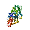 8hfnC 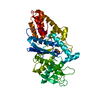 8hfoC 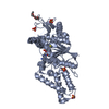 3c8zS S: Starting model for refinement C: citing same article ( |
|---|---|
| Similar structure data |
-Links
 PDBj
PDBj- Assembly
| Deposited unit | 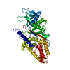
| ||||||||
|---|---|---|---|---|---|---|---|---|---|
| 1 |
| ||||||||
| Unit cell |
|
-Components
| #1: Protein | Mass: 45508.605 Da / Num. of mol.: 1 Source method: isolated from a genetically manipulated source Source: (gene. exp.) Mycolicibacterium smegmatis (bacteria) / Strain: ATCC 700084 / mc(2)155 / Gene: mshC, cysS2, MSMEG_4189, MSMEI_4091 / Plasmid: pETRUK / Production host: References: UniProt: A0QZY0, L-cysteine:1D-myo-inositol 2-amino-2-deoxy-alpha-D-glucopyranoside ligase |
|---|---|
| #2: Chemical | ChemComp-CA /   Mass: 40.078 Da / Num. of mol.: 1 / Source method: obtained synthetically / Formula: Ca Mass: 40.078 Da / Num. of mol.: 1 / Source method: obtained synthetically / Formula: Ca |
| #3: Chemical | ChemComp-ZN /   Mass: 65.409 Da / Num. of mol.: 1 / Source method: obtained synthetically / Formula: Zn Mass: 65.409 Da / Num. of mol.: 1 / Source method: obtained synthetically / Formula: Zn |
| #4: Water | ChemComp-HOH /  Mass: 18.015 Da / Num. of mol.: 65 / Source method: isolated from a natural source / Formula: H2O Mass: 18.015 Da / Num. of mol.: 65 / Source method: isolated from a natural source / Formula: H2O |
| Has ligand of interest | N |
-Experimental details
-Experiment
| Experiment | Method: X-RAY DIFFRACTION / Number of used crystals: 1 |
|---|
- Sample preparation
| Crystal | Density Matthews: 3.16 Å3/Da / Density % sol: 61.07 % |
|---|---|
| Crystal grow | Temperature: 293 K / Method: vapor diffusion, hanging drop / pH: 7.1 Details: Holo protein at 10 mg/mL in 10 mM Tris pH 7, 100 mM NaCl, 2.5 mM 2-mercaptoethanol was mixed with 25 mM CaCl2, 25 mM MgCl2, PEG8000 5-8% (w/v), Morpheus buffer system 2 pH 7.1, ethylene ...Details: Holo protein at 10 mg/mL in 10 mM Tris pH 7, 100 mM NaCl, 2.5 mM 2-mercaptoethanol was mixed with 25 mM CaCl2, 25 mM MgCl2, PEG8000 5-8% (w/v), Morpheus buffer system 2 pH 7.1, ethylene glycol 20% (v/v) in a 1:1 ratio. Suitable crystals were immersed in mother liquor supplemented with 22% (v/v) ethylene glycol as a cryoprotectant and flash frozen in liquid nitrogen for subsequent data collection. |
-Data collection
| Diffraction | Mean temperature: 100 K / Serial crystal experiment: N |
|---|---|
| Diffraction source | Source: SYNCHROTRON / Site: SOLEIL  / Beamline: PROXIMA 1 / Wavelength: 0.911648 Å / Beamline: PROXIMA 1 / Wavelength: 0.911648 Å |
| Detector | Type: DECTRIS EIGER X 16M / Detector: PIXEL / Date: Dec 16, 2019 |
| Radiation | Protocol: SINGLE WAVELENGTH / Monochromatic (M) / Laue (L): M / Scattering type: x-ray |
| Radiation wavelength | Wavelength: 0.911648 Å / Relative weight: 1 |
| Reflection | Resolution: 2.41→69.81 Å / Num. obs: 23600 / % possible obs: 100 % / Redundancy: 26.1 % / CC1/2: 0.999 / Rmerge(I) obs: 0.172 / Rpim(I) all: 0.034 / Rrim(I) all: 0.175 / Χ2: 0.96 / Net I/σ(I): 15.3 / Num. measured all: 617009 |
| Reflection shell | Resolution: 2.41→2.54 Å / % possible obs: 100 % / Redundancy: 26.7 % / Rmerge(I) obs: 2.385 / Num. measured all: 89091 / Num. unique obs: 3341 / CC1/2: 0.778 / Rpim(I) all: 0.468 / Rrim(I) all: 2.431 / Χ2: 0.84 / Net I/σ(I) obs: 1.7 |
- Processing
| Software |
| |||||||||||||||||||||||||||||||||||||||||||||||||||||||||||||||||||||||||||
|---|---|---|---|---|---|---|---|---|---|---|---|---|---|---|---|---|---|---|---|---|---|---|---|---|---|---|---|---|---|---|---|---|---|---|---|---|---|---|---|---|---|---|---|---|---|---|---|---|---|---|---|---|---|---|---|---|---|---|---|---|---|---|---|---|---|---|---|---|---|---|---|---|---|---|---|---|
| Refinement | Method to determine structure: MOLECULAR REPLACEMENT Starting model: 3C8Z Resolution: 2.41→48.32 Å / SU ML: 0.32 / Cross valid method: THROUGHOUT / σ(F): 1.35 / Phase error: 26.15 / Stereochemistry target values: ML
| |||||||||||||||||||||||||||||||||||||||||||||||||||||||||||||||||||||||||||
| Solvent computation | Shrinkage radii: 0.9 Å / VDW probe radii: 1.11 Å / Solvent model: FLAT BULK SOLVENT MODEL | |||||||||||||||||||||||||||||||||||||||||||||||||||||||||||||||||||||||||||
| Refinement step | Cycle: LAST / Resolution: 2.41→48.32 Å
| |||||||||||||||||||||||||||||||||||||||||||||||||||||||||||||||||||||||||||
| Refine LS restraints |
| |||||||||||||||||||||||||||||||||||||||||||||||||||||||||||||||||||||||||||
| LS refinement shell |
| |||||||||||||||||||||||||||||||||||||||||||||||||||||||||||||||||||||||||||
| Refinement TLS params. | Method: refined / Refine-ID: X-RAY DIFFRACTION
| |||||||||||||||||||||||||||||||||||||||||||||||||||||||||||||||||||||||||||
| Refinement TLS group |
|
