 Movie
Movie Controller

[English] 日本語
 Yorodumi
- PDB-8he1: The structure of chitin deacetylase Pst_13661 from Puccinia strii... -
+ Open data

- Basic information
| Entry | Database: PDB / ID: 8he1 | ||||||
|---|---|---|---|---|---|---|---|
| Title | The structure of chitin deacetylase Pst_13661 from Puccinia striiformis f. sp. tritici | ||||||
 Components Components | Chitin deacetylase | ||||||
 Keywords Keywords | HYDROLASE / Chitin deacetylase / Plant pathogenic fungi / Virulence factor | ||||||
| Function / homology |  Function and homology information Function and homology informationhydrolase activity, acting on carbon-nitrogen (but not peptide) bonds / side of membrane / carbohydrate metabolic process / metal ion binding / plasma membrane Similarity search - Function | ||||||
| Biological species |  Puccinia striiformis f. sp. tritici (fungus) Puccinia striiformis f. sp. tritici (fungus) | ||||||
| Method |  X-RAY DIFFRACTION / SYNCHROTRON / MOLECULAR REPLACEMENT / Resolution: 1.61 Å X-RAY DIFFRACTION / SYNCHROTRON / MOLECULAR REPLACEMENT / Resolution: 1.61 Å | ||||||
 Authors Authors | Liu, L. / Li, Y.C. / Zhou, Y. / Yang, Q. | ||||||
| Funding support |  China, 1items China, 1items
| ||||||
 Citation Citation | Journal: Nat Commun / Year: 2023 Title: Inhibition of chitin deacetylases to attenuate plant fungal diseases. Authors: Liu, L. / Xia, Y. / Li, Y. / Zhou, Y. / Su, X. / Yan, X. / Wang, Y. / Liu, W. / Cheng, H. / Wang, Y. / Yang, Q. | ||||||
| History |
|
- Structure visualization
| Structure viewer | Molecule: MolmilJmol/JSmol |
|---|
- Downloads & links
-Download
| PDBx/mmCIF format | 8he1.cif.gz | 70.3 KB | Display | PDBx/mmCIF format |
|---|---|---|---|---|
| PDB format | pdb8he1.ent.gz | 47.9 KB | Display | PDB format |
| PDBx/mmJSON format | 8he1.json.gz | Tree view | PDBx/mmJSON format | |
| Others |  Other downloads Other downloads |
-Validation report
| Arichive directory | https://data.pdbj.org/pub/pdb/validation_reports/he/8he1ftp://data.pdbj.org/pub/pdb/validation_reports/he/8he1 | HTTPS FTP |
|---|
-Related structure data
| Related structure data | 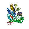 8he2C 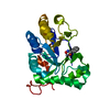 8he4C  8hf9C 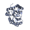 8hfaC  2iw0S S: Starting model for refinement C: citing same article ( |
|---|---|
| Similar structure data |
-Links
 PDBj
PDBj
- Assembly
| Deposited unit | 
| ||||||||||||
|---|---|---|---|---|---|---|---|---|---|---|---|---|---|
| 1 |
| ||||||||||||
| Unit cell |
|
-Components
| #1: Protein | Mass: 31636.381 Da / Num. of mol.: 1 Source method: isolated from a genetically manipulated source Source: (gene. exp.) Puccinia striiformis f. sp. tritici (fungus)Production host: Komagataella pastoris (fungus) / References: UniProt: A0A2S4WL56, chitin deacetylase |
|---|---|
| #2: Chemical | ChemComp-ZN /   Mass: 65.409 Da / Num. of mol.: 1 / Source method: obtained synthetically / Formula: Zn / Feature type: SUBJECT OF INVESTIGATION Mass: 65.409 Da / Num. of mol.: 1 / Source method: obtained synthetically / Formula: Zn / Feature type: SUBJECT OF INVESTIGATION |
| #3: Chemical | ChemComp-BHO / 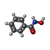  Mass: 137.136 Da / Num. of mol.: 1 / Source method: isolated from a natural source / Formula: C7H7NO2 / Feature type: SUBJECT OF INVESTIGATION Mass: 137.136 Da / Num. of mol.: 1 / Source method: isolated from a natural source / Formula: C7H7NO2 / Feature type: SUBJECT OF INVESTIGATION |
| #4: Water | ChemComp-HOH /  Mass: 18.015 Da / Num. of mol.: 298 / Source method: isolated from a natural source / Formula: H2O Mass: 18.015 Da / Num. of mol.: 298 / Source method: isolated from a natural source / Formula: H2O |
| Has ligand of interest | Y |
| Has protein modification | Y |
-Experimental details
-Experiment
| Experiment | Method: X-RAY DIFFRACTION / Number of used crystals: 1 |
|---|
- Sample preparation
| Crystal | Density Matthews: 2.18 Å3/Da / Density % sol: 43.6 % |
|---|---|
| Crystal grow | Temperature: 277 K / Method: vapor diffusion, hanging drop Details: 1.1M Sodium malonate, 0.1M HEPES pH 7.0, 0.5% v/v Jeffamine ED-2001 |
-Data collection
| Diffraction | Mean temperature: 100 K / Serial crystal experiment: N |
|---|---|
| Diffraction source | Source: SYNCHROTRON / Site: SSRF / Beamline: BL17B1 / Wavelength: 0.97925 Å |
| Detector | Type: DECTRIS PILATUS 2M / Detector: PIXEL / Date: May 29, 2021 |
| Radiation | Protocol: SINGLE WAVELENGTH / Monochromatic (M) / Laue (L): M / Scattering type: x-ray |
| Radiation wavelength | Wavelength: 0.97925 Å / Relative weight: 1 |
| Reflection | Resolution: 1.61→50 Å / Num. obs: 36799 / % possible obs: 99.2 % / Redundancy: 13 % / Biso Wilson estimate: 16.46 Å2 / Rmerge(I) obs: 0.134 / Net I/σ(I): 4.8 |
| Reflection shell | Resolution: 1.61→1.64 Å / Rmerge(I) obs: 0.421 / Num. unique obs: 1731 |
- Processing
| Software |
| |||||||||||||||||||||||||||||||||||||||||||||||||||||||||||||||||||||||||||||||||||||||||||||||||||||||||
|---|---|---|---|---|---|---|---|---|---|---|---|---|---|---|---|---|---|---|---|---|---|---|---|---|---|---|---|---|---|---|---|---|---|---|---|---|---|---|---|---|---|---|---|---|---|---|---|---|---|---|---|---|---|---|---|---|---|---|---|---|---|---|---|---|---|---|---|---|---|---|---|---|---|---|---|---|---|---|---|---|---|---|---|---|---|---|---|---|---|---|---|---|---|---|---|---|---|---|---|---|---|---|---|---|---|---|
| Refinement | Method to determine structure: MOLECULAR REPLACEMENT Starting model: 2iw0 Resolution: 1.61→26.51 Å / SU ML: 0.1456 / Cross valid method: FREE R-VALUE / σ(F): 1.38 / Phase error: 16.5778 Stereochemistry target values: GeoStd + Monomer Library + CDL v1.2
| |||||||||||||||||||||||||||||||||||||||||||||||||||||||||||||||||||||||||||||||||||||||||||||||||||||||||
| Solvent computation | Shrinkage radii: 0.9 Å / VDW probe radii: 1.11 Å / Solvent model: FLAT BULK SOLVENT MODEL | |||||||||||||||||||||||||||||||||||||||||||||||||||||||||||||||||||||||||||||||||||||||||||||||||||||||||
| Displacement parameters | Biso mean: 18.94 Å2 | |||||||||||||||||||||||||||||||||||||||||||||||||||||||||||||||||||||||||||||||||||||||||||||||||||||||||
| Refinement step | Cycle: LAST / Resolution: 1.61→26.51 Å
| |||||||||||||||||||||||||||||||||||||||||||||||||||||||||||||||||||||||||||||||||||||||||||||||||||||||||
| Refine LS restraints |
| |||||||||||||||||||||||||||||||||||||||||||||||||||||||||||||||||||||||||||||||||||||||||||||||||||||||||
| LS refinement shell |
|
