 Movie
Movie Controller

[English] 日本語
 Yorodumi
- PDB-8hdl: Crystal structure of ASFV trans geranylgeranyl diphosphate syntha... -
+ Open data

- Basic information
| Entry | Database: PDB / ID: 8hdl | ||||||
|---|---|---|---|---|---|---|---|
| Title | Crystal structure of ASFV trans geranylgeranyl diphosphate synthase B318L | ||||||
 Components Components | Trans-prenyltransferase | ||||||
 Keywords Keywords | TRANSFERASE / ASFV | ||||||
| Function / homology |  Function and homology information Function and homology informationgeranylfarnesyl diphosphate synthase activity / geranylgeranyl diphosphate biosynthetic process / geranylgeranyl diphosphate synthase / geranyl diphosphate biosynthetic process / Transferases; Transferring alkyl or aryl groups, other than methyl groups / dimethylallyltranstransferase / geranylgeranyl diphosphate synthase activity / (2E,6E)-farnesyl diphosphate synthase / farnesyl diphosphate biosynthetic process / dimethylallyltranstransferase activity ...geranylfarnesyl diphosphate synthase activity / geranylgeranyl diphosphate biosynthetic process / geranylgeranyl diphosphate synthase / geranyl diphosphate biosynthetic process / Transferases; Transferring alkyl or aryl groups, other than methyl groups / dimethylallyltranstransferase / geranylgeranyl diphosphate synthase activity / (2E,6E)-farnesyl diphosphate synthase / farnesyl diphosphate biosynthetic process / dimethylallyltranstransferase activity / (2E,6E)-farnesyl diphosphate synthase activity / host cell endoplasmic reticulum / host cell membrane / metal ion binding / membrane Similarity search - Function | ||||||
| Biological species |   African swine fever virus African swine fever virus | ||||||
| Method |  X-RAY DIFFRACTION / SYNCHROTRON / MOLECULAR REPLACEMENT / Resolution: 3.198 Å X-RAY DIFFRACTION / SYNCHROTRON / MOLECULAR REPLACEMENT / Resolution: 3.198 Å | ||||||
 Authors Authors | Zhao, H.F. | ||||||
| Funding support |  China, 1items China, 1items
| ||||||
 Citation Citation | Journal: Int J Mol Sci / Year: 2023 Title: Exploring AlphaFold2's Performance on Predicting Amino Acid Side-Chain Conformations and Its Utility in Crystal Structure Determination of B318L Protein. Authors: Zhao, H. / Zhang, H. / She, Z. / Gao, Z. / Wang, Q. / Geng, Z. / Dong, Y. | ||||||
| History |
|
- Structure visualization
| Structure viewer | Molecule: MolmilJmol/JSmol |
|---|
- Downloads & links
-Download
| PDBx/mmCIF format | 8hdl.cif.gz | 118.6 KB | Display | PDBx/mmCIF format |
|---|---|---|---|---|
| PDB format | pdb8hdl.ent.gz | 91.4 KB | Display | PDB format |
| PDBx/mmJSON format | 8hdl.json.gz | Tree view | PDBx/mmJSON format | |
| Others |  Other downloads Other downloads |
-Validation report
| Arichive directory | https://data.pdbj.org/pub/pdb/validation_reports/hd/8hdlftp://data.pdbj.org/pub/pdb/validation_reports/hd/8hdl | HTTPS FTP |
|---|
-Related structure data
| Similar structure data |
|---|
-Links
 PDBj
PDBj
- Assembly
| Deposited unit | 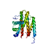
| ||||||||
|---|---|---|---|---|---|---|---|---|---|
| 1 |
| ||||||||
| Unit cell |
|
-Components
| #1: Protein | Mass: 32329.084 Da / Num. of mol.: 1 Source method: isolated from a genetically manipulated source Source: (gene. exp.) African swine fever virus (strain Badajoz 1971 Vero-adapted)Gene: Ba71V-074, B318L / Production host:  References: UniProt: Q65164, Transferases; Transferring alkyl or aryl groups, other than methyl groups, dimethylallyltranstransferase, geranylgeranyl diphosphate synthase, (2E,6E)-farnesyl diphosphate synthase |
|---|
-Experimental details
-Experiment
| Experiment | Method: X-RAY DIFFRACTION / Number of used crystals: 1 |
|---|
- Sample preparation
| Crystal | Density Matthews: 2.65 Å3/Da / Density % sol: 53.57 % |
|---|---|
| Crystal grow | Temperature: 293.15 K / Method: vapor diffusion, sitting drop / Details: 0.1 M HEPES pH 7.5, 4.3 M NaCl |
-Data collection
| Diffraction | Mean temperature: 100 K / Serial crystal experiment: N |
|---|---|
| Diffraction source | Source: SYNCHROTRON / Site: SSRF / Beamline: BL10U2 / Wavelength: 0.9792 Å |
| Detector | Type: DECTRIS EIGER X 16M / Detector: PIXEL / Date: Mar 7, 2022 |
| Radiation | Protocol: SINGLE WAVELENGTH / Monochromatic (M) / Laue (L): M / Scattering type: x-ray |
| Radiation wavelength | Wavelength: 0.9792 Å / Relative weight: 1 |
| Reflection | Resolution: 3.198→48.21 Å / Num. obs: 6579 / % possible obs: 99.7 % / Redundancy: 31.7 % / CC1/2: 0.998 / Rmerge(I) obs: 0.297 / Net I/σ(I): 15.6 |
| Reflection shell | Resolution: 3.2→3.28 Å / Num. unique obs: 472 / CC1/2: 0.718 |
- Processing
| Software |
| ||||||||||||||||||||||||||||||||||||||||||
|---|---|---|---|---|---|---|---|---|---|---|---|---|---|---|---|---|---|---|---|---|---|---|---|---|---|---|---|---|---|---|---|---|---|---|---|---|---|---|---|---|---|---|---|
| Refinement | Method to determine structure: MOLECULAR REPLACEMENT Starting model: AlphaFold2 Resolution: 3.198→48.21 Å / SU ML: 0.47 / Cross valid method: FREE R-VALUE / σ(F): 1.34 / Phase error: 32.34 / Stereochemistry target values: ML
| ||||||||||||||||||||||||||||||||||||||||||
| Solvent computation | Shrinkage radii: 0.9 Å / VDW probe radii: 1.11 Å / Solvent model: FLAT BULK SOLVENT MODEL | ||||||||||||||||||||||||||||||||||||||||||
| Refinement step | Cycle: LAST / Resolution: 3.198→48.21 Å
| ||||||||||||||||||||||||||||||||||||||||||
| Refine LS restraints |
| ||||||||||||||||||||||||||||||||||||||||||
| LS refinement shell |
| ||||||||||||||||||||||||||||||||||||||||||
| Refinement TLS params. | Method: refined / Origin x: 11.0849 Å / Origin y: 14.7343 Å / Origin z: -14.278 Å
| ||||||||||||||||||||||||||||||||||||||||||
| Refinement TLS group | Selection details: all |
