 Movie
Movie Controller


+ Open data

- Basic information
| Entry | Database: PDB / ID: 8hc0 | ||||||
|---|---|---|---|---|---|---|---|
| Title | Crystal structure of the extracellular domains of GPR110 | ||||||
 Components Components | (Adhesion G-protein coupled receptor ...) x 3 | ||||||
 Keywords Keywords | CELL ADHESION / Adhesion GPCR / GPR110 / synaptamide / molecular dynamics simulations | ||||||
| Function / homology |  Function and homology information Function and homology informationenergy reserve metabolic process / regulation of lipid metabolic process / fat cell differentiation / synapse assembly / G protein-coupled receptor activity / memory / neuron projection development / adenylate cyclase-activating G protein-coupled receptor signaling pathway / cytoplasmic vesicle / cell surface receptor signaling pathway ...energy reserve metabolic process / regulation of lipid metabolic process / fat cell differentiation / synapse assembly / G protein-coupled receptor activity / memory / neuron projection development / adenylate cyclase-activating G protein-coupled receptor signaling pathway / cytoplasmic vesicle / cell surface receptor signaling pathway / G protein-coupled receptor signaling pathway / extracellular region / membrane / plasma membrane Similarity search - Function | ||||||
| Biological species |  Homo sapiens (human) Homo sapiens (human) | ||||||
| Method |  X-RAY DIFFRACTION / SYNCHROTRON / MOLECULAR REPLACEMENT / Resolution: 2.9 Å X-RAY DIFFRACTION / SYNCHROTRON / MOLECULAR REPLACEMENT / Resolution: 2.9 Å | ||||||
 Authors Authors | Wang, F.F. / Song, G.J. | ||||||
| Funding support |  China, 1items China, 1items
| ||||||
 Citation Citation | Journal: J.Mol.Biol. / Year: 2023 Title: Crystal Structure of the Extracellular Domains of GPR110. Authors: Wang, F. / Wang, Y. / Qiu, W. / Zhang, Q. / Yang, H. / Song, G. | ||||||
| History |
|
- Structure visualization
| Structure viewer | Molecule: MolmilJmol/JSmol |
|---|
- Downloads & links
-Download
| PDBx/mmCIF format | 8hc0.cif.gz | 190.4 KB | Display | PDBx/mmCIF format |
|---|---|---|---|---|
| PDB format | pdb8hc0.ent.gz | 149.6 KB | Display | PDB format |
| PDBx/mmJSON format | 8hc0.json.gz | Tree view | PDBx/mmJSON format | |
| Others |  Other downloads Other downloads |
-Validation report
| Arichive directory | https://data.pdbj.org/pub/pdb/validation_reports/hc/8hc0ftp://data.pdbj.org/pub/pdb/validation_reports/hc/8hc0 | HTTPS FTP |
|---|
-Related structure data
| Similar structure data |
|---|
-Links
 PDBj
PDBj
- Assembly
| Deposited unit | 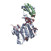
| ||||||||
|---|---|---|---|---|---|---|---|---|---|
| 1 |
| ||||||||
| Unit cell |
|
-Components
-Adhesion G-protein coupled receptor ... , 3 types, 3 molecules ACB
| #1: Protein | Mass: 39499.711 Da / Num. of mol.: 1 Source method: isolated from a genetically manipulated source Source: (gene. exp.) Homo sapiens (human) / Gene: ADGRF1, GPR110, PGR19 / Cell line (production host): HEK293gnt-Production host: Mammalian expression vector Flag-MCS-pcDNA3.1 (others) References: UniProt: Q5T601 |
|---|---|
| #2: Protein/peptide | Mass: 2413.712 Da / Num. of mol.: 1 / Fragment: C-terminal Source method: isolated from a genetically manipulated source Source: (gene. exp.) Homo sapiens (human) / Gene: ADGRF1, GPR110, PGR19 / Cell line (production host): HEK293gnt-Production host: Mammalian expression vector Flag-MCS-pcDNA3.1 (others) References: UniProt: Q5T601 |
| #3: Protein | Mass: 6612.419 Da / Num. of mol.: 1 / Fragment: N-terminal Source method: isolated from a genetically manipulated source Source: (gene. exp.) Homo sapiens (human) / Gene: ADGRF1, GPR110, PGR19 / Cell line (production host): HEK293gnt-Production host: Mammalian expression vector Flag-MCS-pcDNA3.1 (others) References: UniProt: Q5T601 |
-Sugars , 2 types, 8 molecules 
| #4: Polysaccharide | alpha-D-mannopyranose-(1-3)-alpha-D-mannopyranose-(1-6)-[alpha-D-mannopyranose-(1-3)]alpha-D- ...alpha-D-mannopyranose-(1-3)-alpha-D-mannopyranose-(1-6)-[alpha-D-mannopyranose-(1-3)]alpha-D-mannopyranose-(1-4)-2-acetamido-2-deoxy-beta-D-glucopyranose-(1-4)-2-acetamido-2-deoxy-beta-D-glucopyranose |
|---|---|
| #5: Sugar | ChemComp-NAG /  Type: D-saccharide, beta linking / Mass: 221.208 Da / Num. of mol.: 7 Type: D-saccharide, beta linking / Mass: 221.208 Da / Num. of mol.: 7Source method: isolated from a genetically manipulated source Formula: C8H15NO6 / Feature type: SUBJECT OF INVESTIGATION |
-Non-polymers , 1 types, 45 molecules 
| #6: Water | ChemComp-HOH / Mass: 18.015 Da / Num. of mol.: 45 / Source method: isolated from a natural source / Formula: H2O |
|---|
-Details
| Has ligand of interest | Y |
|---|---|
| Has protein modification | Y |
-Experimental details
-Experiment
| Experiment | Method: X-RAY DIFFRACTION / Number of used crystals: 1 |
|---|
- Sample preparation
| Crystal | Density Matthews: 2.35 Å3/Da / Density % sol: 47.65 % |
|---|---|
| Crystal grow | Temperature: 289 K / Method: vapor diffusion Details: 0.2 M ammonium sulfate, 0.1 M sodium acetate trihydrate pH 4.6, 30% PEG-MME 2,000, 15% glycerol |
-Data collection
| Diffraction | Mean temperature: 100 K / Serial crystal experiment: N | ||||||||||||||||||||||||||||||||||||||||||||||||||||||||||||
|---|---|---|---|---|---|---|---|---|---|---|---|---|---|---|---|---|---|---|---|---|---|---|---|---|---|---|---|---|---|---|---|---|---|---|---|---|---|---|---|---|---|---|---|---|---|---|---|---|---|---|---|---|---|---|---|---|---|---|---|---|---|
| Diffraction source | Source: SYNCHROTRON / Site: SSRF / Beamline: BL10U2 / Wavelength: 0.9792 Å | ||||||||||||||||||||||||||||||||||||||||||||||||||||||||||||
| Detector | Type: DECTRIS EIGER X 16M / Detector: PIXEL / Date: Mar 10, 2022 | ||||||||||||||||||||||||||||||||||||||||||||||||||||||||||||
| Radiation | Protocol: SINGLE WAVELENGTH / Monochromatic (M) / Laue (L): M / Scattering type: x-ray | ||||||||||||||||||||||||||||||||||||||||||||||||||||||||||||
| Radiation wavelength | Wavelength: 0.9792 Å / Relative weight: 1 | ||||||||||||||||||||||||||||||||||||||||||||||||||||||||||||
| Reflection | Resolution: 2.9→43.8 Å / Num. obs: 18390 / % possible obs: 98 % / Redundancy: 3.18 % / CC1/2: 0.989 / Rmerge(I) obs: 0.124 / Rrim(I) all: 0.149 / Net I/σ(I): 9.68 | ||||||||||||||||||||||||||||||||||||||||||||||||||||||||||||
| Reflection shell |
|
- Processing
| Software |
| ||||||||||||||||||||||||||||||||||||||||||||||||||||||||||||||||||||||||||||||||||||||||||||||||||||
|---|---|---|---|---|---|---|---|---|---|---|---|---|---|---|---|---|---|---|---|---|---|---|---|---|---|---|---|---|---|---|---|---|---|---|---|---|---|---|---|---|---|---|---|---|---|---|---|---|---|---|---|---|---|---|---|---|---|---|---|---|---|---|---|---|---|---|---|---|---|---|---|---|---|---|---|---|---|---|---|---|---|---|---|---|---|---|---|---|---|---|---|---|---|---|---|---|---|---|---|---|---|
| Refinement | Method to determine structure: MOLECULAR REPLACEMENT Starting model: predicted by AlphaFold2 Resolution: 2.9→19.8 Å / SU ML: 0.44 / Cross valid method: THROUGHOUT / σ(F): 1.38 / Phase error: 30.88 / Stereochemistry target values: ML
| ||||||||||||||||||||||||||||||||||||||||||||||||||||||||||||||||||||||||||||||||||||||||||||||||||||
| Solvent computation | Shrinkage radii: 0.9 Å / VDW probe radii: 1.1 Å / Solvent model: FLAT BULK SOLVENT MODEL | ||||||||||||||||||||||||||||||||||||||||||||||||||||||||||||||||||||||||||||||||||||||||||||||||||||
| Refinement step | Cycle: LAST / Resolution: 2.9→19.8 Å
| ||||||||||||||||||||||||||||||||||||||||||||||||||||||||||||||||||||||||||||||||||||||||||||||||||||
| Refine LS restraints |
| ||||||||||||||||||||||||||||||||||||||||||||||||||||||||||||||||||||||||||||||||||||||||||||||||||||
| LS refinement shell |
| ||||||||||||||||||||||||||||||||||||||||||||||||||||||||||||||||||||||||||||||||||||||||||||||||||||
| Refinement TLS params. | Method: refined / Refine-ID: X-RAY DIFFRACTION
| ||||||||||||||||||||||||||||||||||||||||||||||||||||||||||||||||||||||||||||||||||||||||||||||||||||
| Refinement TLS group |
|
