 Movie
Movie Controller

[English] 日本語
 Yorodumi
- PDB-8haz: Crystal structure of Caenorhabditis elegans NMAD-1 in complex wit... -
+ Open data

- Basic information
| Entry | Database: PDB / ID: 8haz | ||||||
|---|---|---|---|---|---|---|---|
| Title | Crystal structure of Caenorhabditis elegans NMAD-1 in complex with ligand I | ||||||
 Components Components | DNA N6-methyl adenine demethylase | ||||||
 Keywords Keywords | OXIDOREDUCTASE / NMAD-1A / GENE REGULATION | ||||||
| Function / homology |  Function and homology information Function and homology informationpositive regulation of egg-laying behavior / DNA N6-methyladenine demethylase / DNA N6-methyladenine demethylase activity / positive regulation of fertilization / meiotic chromosome condensation / demethylase activity / contractile ring / demethylation / positive regulation of meiosis I / positive regulation of chromosome organization ...positive regulation of egg-laying behavior / DNA N6-methyladenine demethylase / DNA N6-methyladenine demethylase activity / positive regulation of fertilization / meiotic chromosome condensation / demethylase activity / contractile ring / demethylation / positive regulation of meiosis I / positive regulation of chromosome organization / 2-oxoglutarate-dependent dioxygenase activity / actomyosin structure organization / positive regulation of double-strand break repair / midbody / DNA replication / metal ion binding / nucleus Similarity search - Function | ||||||
| Biological species |  | ||||||
| Method |  X-RAY DIFFRACTION / SYNCHROTRON / AB INITIO PHASING / Resolution: 2.66 Å X-RAY DIFFRACTION / SYNCHROTRON / AB INITIO PHASING / Resolution: 2.66 Å | ||||||
 Authors Authors | Shang, G. / Chen, Z. | ||||||
| Funding support |  China, 1items China, 1items
| ||||||
 Citation Citation | Journal: Int J Mol Sci / Year: 2024 Title: Structural Basis of Nucleic Acid Recognition and 6mA Demethylation by Caenorhabditis elegans NMAD-1A. Authors: Shang, G. / Yang, M. / Li, M. / Ma, L. / Liu, Y. / Ma, J. / Chen, Y. / Wang, X. / Fan, S. / Xie, M. / Wu, W. / Dai, S. / Chen, Z. | ||||||
| History |
|
- Structure visualization
| Structure viewer | Molecule: MolmilJmol/JSmol |
|---|
- Downloads & links
-Download
| PDBx/mmCIF format | 8haz.cif.gz | 54.7 KB | Display | PDBx/mmCIF format |
|---|---|---|---|---|
| PDB format | pdb8haz.ent.gz | 36.4 KB | Display | PDB format |
| PDBx/mmJSON format | 8haz.json.gz | Tree view | PDBx/mmJSON format | |
| Others |  Other downloads Other downloads |
-Validation report
| Summary document | 8haz_validation.pdf.gz | 873.1 KB | Display | wwPDB validaton report |
|---|---|---|---|---|
| Full document | 8haz_full_validation.pdf.gz | 873.3 KB | Display | |
| Data in XML | 8haz_validation.xml.gz | 10 KB | Display | |
| Data in CIF | 8haz_validation.cif.gz | 13.1 KB | Display | |
| Arichive directory | https://data.pdbj.org/pub/pdb/validation_reports/ha/8hazftp://data.pdbj.org/pub/pdb/validation_reports/ha/8haz | HTTPS FTP |
-Related structure data


-Links
 PDBj
PDBj- Assembly
| Deposited unit | 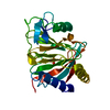
| ||||||||
|---|---|---|---|---|---|---|---|---|---|
| 1 |
| ||||||||
| Unit cell |
|
-Components
| #1: Protein | Mass: 28516.553 Da / Num. of mol.: 1 Source method: isolated from a genetically manipulated source Source: (gene. exp.)  prokaryote coculture (bacteria) prokaryote coculture (bacteria)References: UniProt: Q8MNT9, DNA N6-methyladenine demethylase |
|---|---|
| #2: Chemical | ChemComp-SO4 /   Mass: 96.063 Da / Num. of mol.: 1 / Source method: obtained synthetically / Formula: SO4 / Feature type: SUBJECT OF INVESTIGATION Mass: 96.063 Da / Num. of mol.: 1 / Source method: obtained synthetically / Formula: SO4 / Feature type: SUBJECT OF INVESTIGATION |
| #3: Water | ChemComp-HOH /  Mass: 18.015 Da / Num. of mol.: 73 / Source method: isolated from a natural source / Formula: H2O Mass: 18.015 Da / Num. of mol.: 73 / Source method: isolated from a natural source / Formula: H2O |
| Has ligand of interest | Y |
-Experimental details
-Experiment
| Experiment | Method: X-RAY DIFFRACTION / Number of used crystals: 1 |
|---|
- Sample preparation
| Crystal | Density Matthews: 1.91 Å3/Da / Density % sol: 35.56 % |
|---|---|
| Crystal grow | Temperature: 289 K / Method: vapor diffusion, sitting drop / pH: 6.5 Details: 25% PEG 3350, 200 mM, CH3COONH4, 100 mM Bis-Tris pH 6.5 |
-Data collection
| Diffraction | Mean temperature: 293 K / Serial crystal experiment: N |
|---|---|
| Diffraction source | Source: SYNCHROTRON / Site: SSRF / Beamline: BL02U1 / Wavelength: 0.9793 Å |
| Detector | Type: DECTRIS EIGER X 16M / Detector: PIXEL / Date: Oct 6, 2021 |
| Radiation | Protocol: SINGLE WAVELENGTH / Monochromatic (M) / Laue (L): M / Scattering type: x-ray |
| Radiation wavelength | Wavelength: 0.9793 Å / Relative weight: 1 |
| Reflection | Resolution: 2.65→50 Å / Num. obs: 6106 / % possible obs: 90.3 % / Redundancy: 3.4 % / CC1/2: 0.965 / Net I/σ(I): 9.7 |
| Reflection shell | Resolution: 2.65→2.7 Å / Num. unique obs: 597 / CC1/2: 0.894 |
- Processing
| Software |
| ||||||||||||||||||||||||
|---|---|---|---|---|---|---|---|---|---|---|---|---|---|---|---|---|---|---|---|---|---|---|---|---|---|
| Refinement | Method to determine structure: AB INITIO PHASING / Resolution: 2.66→37.54 Å / SU ML: 0.36 / Cross valid method: FREE R-VALUE / σ(F): 1.38 / Phase error: 25.92 / Stereochemistry target values: ML
| ||||||||||||||||||||||||
| Solvent computation | Shrinkage radii: 0.9 Å / VDW probe radii: 1.11 Å / Solvent model: FLAT BULK SOLVENT MODEL | ||||||||||||||||||||||||
| Refinement step | Cycle: LAST / Resolution: 2.66→37.54 Å
| ||||||||||||||||||||||||
| Refine LS restraints |
| ||||||||||||||||||||||||
| LS refinement shell |
|
