 Movie
Movie Controller

[English] 日本語
 Yorodumi
- PDB-8h91: Crystal structure of SARS-CoV-2 spike receptor-binding domain in ... -
+ Open data

- Basic information
| Entry | Database: PDB / ID: 8h91 | ||||||
|---|---|---|---|---|---|---|---|
| Title | Crystal structure of SARS-CoV-2 spike receptor-binding domain in complex with nanobody N19 | ||||||
 Components Components |
| ||||||
 Keywords Keywords | VIRAL PROTEIN/IMMUNE SYSTEM / SARS-CoV-2 / Spike receptor-binding domain nanobody / VIRAL PROTEIN / VIRAL PROTEIN-IMMUNE SYSTEM complex | ||||||
| Function / homology |  Function and homology information Function and homology informationMaturation of spike protein / viral translation / Translation of Structural Proteins / Virion Assembly and Release / host cell surface / host extracellular space / suppression by virus of host tetherin activity / Induction of Cell-Cell Fusion / structural constituent of virion / entry receptor-mediated virion attachment to host cell ...Maturation of spike protein / viral translation / Translation of Structural Proteins / Virion Assembly and Release / host cell surface / host extracellular space / suppression by virus of host tetherin activity / Induction of Cell-Cell Fusion / structural constituent of virion / entry receptor-mediated virion attachment to host cell / host cell endoplasmic reticulum-Golgi intermediate compartment membrane / receptor-mediated endocytosis of virus by host cell / membrane fusion / Attachment and Entry / positive regulation of viral entry into host cell / receptor-mediated virion attachment to host cell / receptor ligand activity / host cell surface receptor binding / fusion of virus membrane with host plasma membrane / fusion of virus membrane with host endosome membrane / viral envelope / virion attachment to host cell / SARS-CoV-2 activates/modulates innate and adaptive immune responses / host cell plasma membrane / virion membrane / identical protein binding / membrane / plasma membrane Similarity search - Function | ||||||
| Biological species |   Severe acute respiratory syndrome coronavirus 2 Severe acute respiratory syndrome coronavirus 2 | ||||||
| Method |  X-RAY DIFFRACTION / SYNCHROTRON / MOLECULAR REPLACEMENT / Resolution: 3.07 Å X-RAY DIFFRACTION / SYNCHROTRON / MOLECULAR REPLACEMENT / Resolution: 3.07 Å | ||||||
 Authors Authors | Zhang, Y.T. / Li, J. / Zhang, J. | ||||||
| Funding support | 1items
| ||||||
 Citation Citation | Journal: To Be Published Title: Crystal structure of SARS-CoV-2 spike receptor-binding domain in complex with nanobody N19 Authors: Zhang, Y.T. / Li, J. / Zhang, J. | ||||||
| History |
|
- Structure visualization
| Structure viewer | Molecule: MolmilJmol/JSmol |
|---|
- Downloads & links
-Download
| PDBx/mmCIF format | 8h91.cif.gz | 157.6 KB | Display | PDBx/mmCIF format |
|---|---|---|---|---|
| PDB format | pdb8h91.ent.gz | 99.5 KB | Display | PDB format |
| PDBx/mmJSON format | 8h91.json.gz | Tree view | PDBx/mmJSON format | |
| Others |  Other downloads Other downloads |
-Validation report
| Summary document | 8h91_validation.pdf.gz | 444.4 KB | Display | wwPDB validaton report |
|---|---|---|---|---|
| Full document | 8h91_full_validation.pdf.gz | 452.7 KB | Display | |
| Data in XML | 8h91_validation.xml.gz | 22.9 KB | Display | |
| Data in CIF | 8h91_validation.cif.gz | 31 KB | Display | |
| Arichive directory | https://data.pdbj.org/pub/pdb/validation_reports/h9/8h91ftp://data.pdbj.org/pub/pdb/validation_reports/h9/8h91 | HTTPS FTP |
-Related structure data
| Related structure data |  7w1sS S: Starting model for refinement |
|---|---|
| Similar structure data |
-Links
 PDBj
PDBj


- Assembly
| Deposited unit | 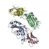
| ||||||||||||
|---|---|---|---|---|---|---|---|---|---|---|---|---|---|
| 1 |
| ||||||||||||
| Unit cell |
|
-Components
| #1: Protein | Mass: 21772.391 Da / Num. of mol.: 2 Source method: isolated from a genetically manipulated source Source: (gene. exp.) Severe acute respiratory syndrome coronavirus 2Gene: S, 2 / Production host:   Spodoptera frugiperda (fall armyworm) / References: UniProt: P0DTC2 Spodoptera frugiperda (fall armyworm) / References: UniProt: P0DTC2#2: Antibody | Mass: 12568.915 Da / Num. of mol.: 2 Source method: isolated from a genetically manipulated source Source: (gene. exp.)  |
|---|
-Experimental details
-Experiment
| Experiment | Method: X-RAY DIFFRACTION / Number of used crystals: 1 |
|---|
- Sample preparation
| Crystal | Density Matthews: 2.66 Å3/Da / Density % sol: 53.84 % |
|---|---|
| Crystal grow | Temperature: 298 K / Method: vapor diffusion, hanging drop Details: 1%(w/v) Tryptone, 0.001 M Sodiumazide, 0.05 M HepesSodium pH7.0, 20%(w/v) polyethyleneglycol3350 |
-Data collection
| Diffraction | Mean temperature: 100 K / Serial crystal experiment: N |
|---|---|
| Diffraction source | Source: SYNCHROTRON / Site: SSRF  / Beamline: BL17U1 / Wavelength: 0.979183 Å / Beamline: BL17U1 / Wavelength: 0.979183 Å |
| Detector | Type: ADSC QUANTUM 315r / Detector: CCD / Date: Sep 18, 2021 |
| Radiation | Protocol: SINGLE WAVELENGTH / Monochromatic (M) / Laue (L): M / Scattering type: x-ray |
| Radiation wavelength | Wavelength: 0.979183 Å / Relative weight: 1 |
| Reflection | Resolution: 3.07→55.55 Å / Num. obs: 14976 / % possible obs: 98.9 % / Redundancy: 4.4 % / Biso Wilson estimate: 56.998 Å2 / Rmerge(I) obs: 0.054 / Net I/σ(I): 7.6 |
| Reflection shell | Resolution: 3.07→3.23 Å / Rmerge(I) obs: 1.413 / Num. unique obs: 2124 |
- Processing
| Software |
| |||||||||||||||||||||||||||||||||||
|---|---|---|---|---|---|---|---|---|---|---|---|---|---|---|---|---|---|---|---|---|---|---|---|---|---|---|---|---|---|---|---|---|---|---|---|---|
| Refinement | Method to determine structure: MOLECULAR REPLACEMENT Starting model: 7W1S Resolution: 3.07→55.54 Å / SU ML: 0.463565732501 / Cross valid method: THROUGHOUT / σ(F): 1.338 / Phase error: 27.5093856822 Stereochemistry target values: GeoStd + Monomer Library + CDL v1.2
| |||||||||||||||||||||||||||||||||||
| Solvent computation | Shrinkage radii: 0.9 Å / VDW probe radii: 1.11 Å / Solvent model: FLAT BULK SOLVENT MODEL | |||||||||||||||||||||||||||||||||||
| Displacement parameters | Biso mean: 43.5462081597 Å2 | |||||||||||||||||||||||||||||||||||
| Refinement step | Cycle: LAST / Resolution: 3.07→55.54 Å
| |||||||||||||||||||||||||||||||||||
| Refine LS restraints |
| |||||||||||||||||||||||||||||||||||
| LS refinement shell |
|
