 Movie
Movie Controller

[English] 日本語
 Yorodumi
- PDB-8h79: The crystal structure of cyanorhodopsin-II (CyR-II) P7104R from N... -
+ Open data

- Basic information
| Entry | Database: PDB / ID: 8h79 | |||||||||
|---|---|---|---|---|---|---|---|---|---|---|
| Title | The crystal structure of cyanorhodopsin-II (CyR-II) P7104R from Nodosilinea nodulosa PCC 7104 | |||||||||
 Components Components | cyanorhodopsin-II (CyR-II) P7104R | |||||||||
 Keywords Keywords | MEMBRANE PROTEIN / RETINAL CELL-FREE SYNTHESIS Bacterial type rhodopsin Cyanobacteria | |||||||||
| Function / homology | TETRADECANE / HEXANE / N-OCTANE / HEXADECANE / RETINAL Function and homology information Function and homology information | |||||||||
| Biological species |  Nodosilinea nodulosa PCC 7104 (bacteria) Nodosilinea nodulosa PCC 7104 (bacteria) | |||||||||
| Method |  X-RAY DIFFRACTION / SYNCHROTRON / MOLECULAR REPLACEMENT / Resolution: 2.07 Å X-RAY DIFFRACTION / SYNCHROTRON / MOLECULAR REPLACEMENT / Resolution: 2.07 Å | |||||||||
 Authors Authors | Hosaka, T. / Kimura-Someya, T. / Shirouzu, M. | |||||||||
| Funding support |  Japan, 2items Japan, 2items
| |||||||||
 Citation Citation | Journal: Isme J / Year: 2024 Title: Cyanorhodopsin-II represents a yellow-absorbing proton-pumping rhodopsin clade within cyanobacteria. Authors: Hasegawa-Takano, M. / Hosaka, T. / Kojima, K. / Nishimura, Y. / Kurihara, M. / Nakajima, Y. / Ishizuka-Katsura, Y. / Kimura-Someya, T. / Shirouzu, M. / Sudo, Y. / Yoshizawa, S. | |||||||||
| History |
|
- Structure visualization
| Structure viewer | Molecule: MolmilJmol/JSmol |
|---|
- Downloads & links
-Download
| PDBx/mmCIF format | 8h79.cif.gz | 67.8 KB | Display | PDBx/mmCIF format |
|---|---|---|---|---|
| PDB format | pdb8h79.ent.gz | 46.1 KB | Display | PDB format |
| PDBx/mmJSON format | 8h79.json.gz | Tree view | PDBx/mmJSON format | |
| Others |  Other downloads Other downloads |
-Validation report
| Arichive directory | https://data.pdbj.org/pub/pdb/validation_reports/h7/8h79ftp://data.pdbj.org/pub/pdb/validation_reports/h7/8h79 | HTTPS FTP |
|---|
-Related structure data
| Related structure data | 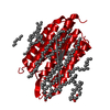 1c3wS S: Starting model for refinement |
|---|---|
| Similar structure data |
-Links
 PDBj
PDBj


- Assembly
| Deposited unit | 
| ||||||||
|---|---|---|---|---|---|---|---|---|---|
| 1 | 
| ||||||||
| Unit cell |
| ||||||||
| Details | There is no clear evidence that the assembly should be a monomer or trimer. |
-Components
-Protein , 1 types, 1 molecules A
| #1: Protein | Mass: 28417.238 Da / Num. of mol.: 1 Source method: isolated from a genetically manipulated source Details: Sequence has been deposited to Genbank with accession number WP_017301364.1 Source: (gene. exp.) Nodosilinea nodulosa PCC 7104 (bacteria)Production host: |
|---|
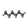
-Non-polymers , 9 types, 59 molecules 


| #2: Chemical | ChemComp-RET /  Mass: 284.436 Da / Num. of mol.: 1 / Source method: obtained synthetically / Formula: C20H28O Mass: 284.436 Da / Num. of mol.: 1 / Source method: obtained synthetically / Formula: C20H28O | ||||||||||||
|---|---|---|---|---|---|---|---|---|---|---|---|---|---|
| #3: Chemical | ChemComp-PG4 /  Mass: 194.226 Da / Num. of mol.: 1 / Source method: obtained synthetically / Formula: C8H18O5 / Comment: precipitant*YM Mass: 194.226 Da / Num. of mol.: 1 / Source method: obtained synthetically / Formula: C8H18O5 / Comment: precipitant*YM | ||||||||||||
| #4: Chemical |  Mass: 86.175 Da / Num. of mol.: 3 / Source method: obtained synthetically / Formula: C6H14 Mass: 86.175 Da / Num. of mol.: 3 / Source method: obtained synthetically / Formula: C6H14#5: Chemical |  Mass: 114.229 Da / Num. of mol.: 3 / Source method: obtained synthetically / Formula: C8H18 Mass: 114.229 Da / Num. of mol.: 3 / Source method: obtained synthetically / Formula: C8H18#6: Chemical |  Mass: 198.388 Da / Num. of mol.: 2 / Source method: obtained synthetically / Formula: C14H30 Mass: 198.388 Da / Num. of mol.: 2 / Source method: obtained synthetically / Formula: C14H30#7: Chemical | ChemComp-R16 / |  Mass: 226.441 Da / Num. of mol.: 1 / Source method: obtained synthetically / Formula: C16H34 Mass: 226.441 Da / Num. of mol.: 1 / Source method: obtained synthetically / Formula: C16H34#8: Chemical | ChemComp-SO4 / |  Mass: 96.063 Da / Num. of mol.: 1 / Source method: obtained synthetically / Formula: SO4 Mass: 96.063 Da / Num. of mol.: 1 / Source method: obtained synthetically / Formula: SO4#9: Chemical | ChemComp-CL / |  Mass: 35.453 Da / Num. of mol.: 1 / Source method: isolated from a natural source / Formula: Cl Mass: 35.453 Da / Num. of mol.: 1 / Source method: isolated from a natural source / Formula: Cl#10: Water | ChemComp-HOH / | Mass: 18.015 Da / Num. of mol.: 46 / Source method: isolated from a natural source / Formula: H2O |
-Details
| Has ligand of interest | N |
|---|---|
| Has protein modification | Y |
-Experimental details
-Experiment
| Experiment | Method: X-RAY DIFFRACTION / Number of used crystals: 1 |
|---|
- Sample preparation
| Crystal | Density Matthews: 2.39 Å3/Da / Density % sol: 48.43 % |
|---|---|
| Crystal grow | Temperature: 293 K / Method: lipidic cubic phase / pH: 6.8 / Details: 100mM HEPES, 46% PEG 400, 400mM lithium sulfate |
-Data collection
| Diffraction | Mean temperature: 93 K / Serial crystal experiment: N | ||||||||||||||||||||||||||||||||||||||||||||||||||||||||||||||||||||||||||||||||||||||||||||||||||||
|---|---|---|---|---|---|---|---|---|---|---|---|---|---|---|---|---|---|---|---|---|---|---|---|---|---|---|---|---|---|---|---|---|---|---|---|---|---|---|---|---|---|---|---|---|---|---|---|---|---|---|---|---|---|---|---|---|---|---|---|---|---|---|---|---|---|---|---|---|---|---|---|---|---|---|---|---|---|---|---|---|---|---|---|---|---|---|---|---|---|---|---|---|---|---|---|---|---|---|---|---|---|
| Diffraction source | Source: SYNCHROTRON / Site: SPring-8 / Beamline: BL32XU / Wavelength: 1 Å | ||||||||||||||||||||||||||||||||||||||||||||||||||||||||||||||||||||||||||||||||||||||||||||||||||||
| Detector | Type: DECTRIS EIGER X 9M / Detector: PIXEL / Date: Jul 2, 2020 | ||||||||||||||||||||||||||||||||||||||||||||||||||||||||||||||||||||||||||||||||||||||||||||||||||||
| Radiation | Protocol: SINGLE WAVELENGTH / Monochromatic (M) / Laue (L): M / Scattering type: x-ray | ||||||||||||||||||||||||||||||||||||||||||||||||||||||||||||||||||||||||||||||||||||||||||||||||||||
| Radiation wavelength | Wavelength: 1 Å / Relative weight: 1 | ||||||||||||||||||||||||||||||||||||||||||||||||||||||||||||||||||||||||||||||||||||||||||||||||||||
| Reflection | Resolution: 2.07→49.6 Å / Num. obs: 17190 / % possible obs: 99.8 % / Redundancy: 12.407 % / Biso Wilson estimate: 27.86 Å2 / CC1/2: 0.997 / Rmerge(I) obs: 0.227 / Rrim(I) all: 0.236 / Χ2: 0.97 / Net I/σ(I): 10.25 / Num. measured all: 213269 / Scaling rejects: 495 | ||||||||||||||||||||||||||||||||||||||||||||||||||||||||||||||||||||||||||||||||||||||||||||||||||||
| Reflection shell | Diffraction-ID: 1
|
- Processing
| Software |
| |||||||||||||||||||||||||||||||||||||||||||||||||||||||||||||||||||||||||||||||||||||||||||
|---|---|---|---|---|---|---|---|---|---|---|---|---|---|---|---|---|---|---|---|---|---|---|---|---|---|---|---|---|---|---|---|---|---|---|---|---|---|---|---|---|---|---|---|---|---|---|---|---|---|---|---|---|---|---|---|---|---|---|---|---|---|---|---|---|---|---|---|---|---|---|---|---|---|---|---|---|---|---|---|---|---|---|---|---|---|---|---|---|---|---|---|---|
| Refinement | Method to determine structure: MOLECULAR REPLACEMENT Starting model: 1C3W Resolution: 2.07→49.6 Å / SU ML: 0.2 / Cross valid method: THROUGHOUT / σ(F): 1.36 / Phase error: 24.66 / Stereochemistry target values: ML
| |||||||||||||||||||||||||||||||||||||||||||||||||||||||||||||||||||||||||||||||||||||||||||
| Solvent computation | Shrinkage radii: 0.9 Å / VDW probe radii: 1.11 Å / Solvent model: FLAT BULK SOLVENT MODEL | |||||||||||||||||||||||||||||||||||||||||||||||||||||||||||||||||||||||||||||||||||||||||||
| Displacement parameters | Biso max: 78.47 Å2 / Biso mean: 29.5547 Å2 / Biso min: 13.85 Å2 | |||||||||||||||||||||||||||||||||||||||||||||||||||||||||||||||||||||||||||||||||||||||||||
| Refinement step | Cycle: final / Resolution: 2.07→49.6 Å
| |||||||||||||||||||||||||||||||||||||||||||||||||||||||||||||||||||||||||||||||||||||||||||
| LS refinement shell | Refine-ID: X-RAY DIFFRACTION / Rfactor Rfree error: 0 / Total num. of bins used: 12
|
