 Movie
Movie Controller

[English] 日本語
 Yorodumi
- PDB-8h2d: The hypothetical protein from Mycobacterium tuberculosis mutant - E47A -
+ Open data

- Basic information
| Entry | Database: PDB / ID: 8h2d | ||||||
|---|---|---|---|---|---|---|---|
| Title | The hypothetical protein from Mycobacterium tuberculosis mutant - E47A | ||||||
 Components Components | Uncharacterized protein Rv1546 | ||||||
 Keywords Keywords | HYDROLASE / RNase | ||||||
| Function / homology | Polyketide cyclase/dehydrase / Polyketide cyclase / dehydrase and lipid transport / START domain / Alpha-D-Glucose-1,6-Bisphosphate; Chain A, domain 4 / START-like domain superfamily / 2-Layer Sandwich / plasma membrane / Alpha Beta / Uncharacterized protein Rv1546 Function and homology information Function and homology information | ||||||
| Biological species |  Mycobacterium tuberculosis H37Rv (bacteria) Mycobacterium tuberculosis H37Rv (bacteria) | ||||||
| Method |  X-RAY DIFFRACTION / SYNCHROTRON / MOLECULAR REPLACEMENT / Resolution: 2.9 Å X-RAY DIFFRACTION / SYNCHROTRON / MOLECULAR REPLACEMENT / Resolution: 2.9 Å | ||||||
 Authors Authors | Kim, D.H. / Na, Y. / Lee, B.J. | ||||||
| Funding support |  Korea, Republic Of, 1items Korea, Republic Of, 1items
| ||||||
 Citation Citation | Journal: Protein Sci. / Year: 2023 Title: Domain swapping of the C-terminal helix promotes the dimerization of a novel ribonuclease protein from Mycobacterium tuberculosis. Authors: Kim, D.H. / Na, Y. / Chang, H. / Boo, J.H. / Kang, S.M. / Jin, C. / Kang, S.J. / Lee, S.Y. / Lee, B.J. | ||||||
| History |
|
- Structure visualization
| Structure viewer | Molecule: MolmilJmol/JSmol |
|---|
- Downloads & links
-Download
| PDBx/mmCIF format | 8h2d.cif.gz | 78.3 KB | Display | PDBx/mmCIF format |
|---|---|---|---|---|
| PDB format | pdb8h2d.ent.gz | 47.1 KB | Display | PDB format |
| PDBx/mmJSON format | 8h2d.json.gz | Tree view | PDBx/mmJSON format | |
| Others |  Other downloads Other downloads |
-Validation report
| Arichive directory | https://data.pdbj.org/pub/pdb/validation_reports/h2/8h2dftp://data.pdbj.org/pub/pdb/validation_reports/h2/8h2d | HTTPS FTP |
|---|
-Related structure data
| Related structure data |  8h0hSC S: Starting model for refinement C: citing same article ( |
|---|---|
| Similar structure data |
-Links
 PDBj
PDBj- Assembly
| Deposited unit | 
| ||||||||||||
|---|---|---|---|---|---|---|---|---|---|---|---|---|---|
| 1 | 
| ||||||||||||
| 2 | 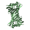
| ||||||||||||
| Unit cell |
|
-Components
| #1: Protein | Mass: 15497.699 Da / Num. of mol.: 2 / Mutation: E47A Source method: isolated from a genetically manipulated source Source: (gene. exp.) Mycobacterium tuberculosis H37Rv (bacteria)Strain: ATCC 25618 / H37Rv / Gene: Rv1546, MTCY48.19c / Production host: #2: Water | ChemComp-HOH / |  Mass: 18.015 Da / Num. of mol.: 18 / Source method: isolated from a natural source / Formula: H2O Mass: 18.015 Da / Num. of mol.: 18 / Source method: isolated from a natural source / Formula: H2OHas protein modification | N | |
|---|
-Experimental details
-Experiment
| Experiment | Method: X-RAY DIFFRACTION / Number of used crystals: 1 |
|---|
- Sample preparation
| Crystal | Density Matthews: 2.17 Å3/Da / Density % sol: 43.39 % |
|---|---|
| Crystal grow | Temperature: 297 K / Method: vapor diffusion, sitting drop Details: 0.1 M Tris, pH 8.5, 0.2 M ammonium sulfate and 25% (w/v) PEG 3350 |
-Data collection
| Diffraction | Mean temperature: 100 K / Serial crystal experiment: N |
|---|---|
| Diffraction source | Source: SYNCHROTRON / Site: PAL/PLS / Beamline: 5C (4A) / Wavelength: 1 Å |
| Detector | Type: DECTRIS EIGER X 9M / Detector: PIXEL / Date: Apr 15, 2021 |
| Radiation | Protocol: SINGLE WAVELENGTH / Monochromatic (M) / Laue (L): M / Scattering type: x-ray |
| Radiation wavelength | Wavelength: 1 Å / Relative weight: 1 |
| Reflection | Resolution: 2.9→50 Å / Num. obs: 6247 / % possible obs: 99.9 % / Redundancy: 12.81 % / Biso Wilson estimate: 87.06 Å2 / CC1/2: 0.999 / Net I/σ(I): 19.79 |
| Reflection shell | Resolution: 2.9→3.07 Å / Num. unique obs: 970 / CC1/2: 0.886 |
- Processing
| Software |
| ||||||||||||||||||||||||
|---|---|---|---|---|---|---|---|---|---|---|---|---|---|---|---|---|---|---|---|---|---|---|---|---|---|
| Refinement | Method to determine structure: MOLECULAR REPLACEMENT Starting model: 8H0H Resolution: 2.9→33.86 Å / SU ML: 0.2497 / Cross valid method: FREE R-VALUE / σ(F): 1.36 / Phase error: 31.3853 Stereochemistry target values: GeoStd + Monomer Library + CDL v1.2
| ||||||||||||||||||||||||
| Solvent computation | Shrinkage radii: 0.9 Å / VDW probe radii: 1.11 Å / Solvent model: FLAT BULK SOLVENT MODEL | ||||||||||||||||||||||||
| Displacement parameters | Biso mean: 82.81 Å2 | ||||||||||||||||||||||||
| Refinement step | Cycle: LAST / Resolution: 2.9→33.86 Å
| ||||||||||||||||||||||||
| Refine LS restraints |
| ||||||||||||||||||||||||
| LS refinement shell |
|
