 Movie
Movie Controller

[English] 日本語
 Yorodumi
- PDB-8h2a: Crystal structure of alcohol dehydrogenase from Formosa agariphila -
+ Open data

- Basic information
| Entry | Database: PDB / ID: 8h2a | ||||||
|---|---|---|---|---|---|---|---|
| Title | Crystal structure of alcohol dehydrogenase from Formosa agariphila | ||||||
 Components Components | Alcohol dehydrogenase | ||||||
 Keywords Keywords | OXIDOREDUCTASE / Alcohol dehydrogenase | ||||||
| Function / homology |  Function and homology information Function and homology information: / S-(hydroxymethyl)glutathione dehydrogenase [NAD(P)+] activity / formaldehyde catabolic process / alcohol dehydrogenase / nucleotide binding / zinc ion binding / cytosol Similarity search - Function | ||||||
| Biological species |  Formosa agariphila (bacteria) Formosa agariphila (bacteria) | ||||||
| Method |  X-RAY DIFFRACTION / SYNCHROTRON / MOLECULAR REPLACEMENT / Resolution: 2.5 Å X-RAY DIFFRACTION / SYNCHROTRON / MOLECULAR REPLACEMENT / Resolution: 2.5 Å | ||||||
 Authors Authors | Brott, S. / Bornscheuer, U.T. / Nam, K.H. | ||||||
| Funding support | 1items
| ||||||
 Citation Citation | Journal: Appl.Microbiol.Biotechnol. / Year: 2023 Title: Unique alcohol dehydrogenases involved in algal sugar utilization by marine bacteria Authors: Brott, S. / Nam, K.H. / Thomas, F. / Dutschei, T. / Reisky, L. / Behrens, M. / Grimm, H.C. / Michel, G. / Schweder, T. / Bornscheuer, U.T. | ||||||
| History |
|
- Structure visualization
| Structure viewer | Molecule: MolmilJmol/JSmol |
|---|
- Downloads & links
-Download
| PDBx/mmCIF format | 8h2a.cif.gz | 546.5 KB | Display | PDBx/mmCIF format |
|---|---|---|---|---|
| PDB format | pdb8h2a.ent.gz | 452.8 KB | Display | PDB format |
| PDBx/mmJSON format | 8h2a.json.gz | Tree view | PDBx/mmJSON format | |
| Others |  Other downloads Other downloads |
-Validation report
| Arichive directory | https://data.pdbj.org/pub/pdb/validation_reports/h2/8h2aftp://data.pdbj.org/pub/pdb/validation_reports/h2/8h2a | HTTPS FTP |
|---|
-Related structure data
| Related structure data | 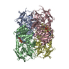 8h2bC 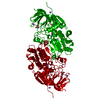 6ljhS S: Starting model for refinement C: citing same article ( |
|---|---|
| Similar structure data |
-Links
 PDBj
PDBj

- Assembly
| Deposited unit | 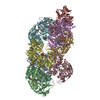
| ||||||||
|---|---|---|---|---|---|---|---|---|---|
| 1 | 
| ||||||||
| 2 | 
| ||||||||
| Unit cell |
|
-Components
| #1: Protein | Mass: 39490.020 Da / Num. of mol.: 8 Source method: isolated from a genetically manipulated source Source: (gene. exp.) Formosa agariphila (bacteria)Strain: DSM 15362 / KCTC 12365 / LMG 23005 / KMM 3901 / M-2Alg 35-1 Gene: BN863_21030 / Production host: #2: Chemical | ChemComp-NAD /   Mass: 663.425 Da / Num. of mol.: 8 / Source method: obtained synthetically / Formula: C21H27N7O14P2 / Feature type: SUBJECT OF INVESTIGATION / Comment: NAD*YM Mass: 663.425 Da / Num. of mol.: 8 / Source method: obtained synthetically / Formula: C21H27N7O14P2 / Feature type: SUBJECT OF INVESTIGATION / Comment: NAD*YM#3: Chemical | ChemComp-ZN /   Mass: 65.409 Da / Num. of mol.: 16 / Source method: obtained synthetically / Formula: Zn / Feature type: SUBJECT OF INVESTIGATION Mass: 65.409 Da / Num. of mol.: 16 / Source method: obtained synthetically / Formula: Zn / Feature type: SUBJECT OF INVESTIGATION#4: Water | ChemComp-HOH / |  Mass: 18.015 Da / Num. of mol.: 288 / Source method: isolated from a natural source / Formula: H2O Mass: 18.015 Da / Num. of mol.: 288 / Source method: isolated from a natural source / Formula: H2OHas ligand of interest | Y | Has protein modification | N | |
|---|
-Experimental details
-Experiment
| Experiment | Method: X-RAY DIFFRACTION / Number of used crystals: 1 |
|---|
- Sample preparation
| Crystal | Density Matthews: 2.35 Å3/Da / Density % sol: 47.63 % |
|---|---|
| Crystal grow | Temperature: 295 K / Method: vapor diffusion, hanging drop / Details: Tris-HCl, pH 7.5, KCl and PEG 3350 |
-Data collection
| Diffraction | Mean temperature: 100 K / Serial crystal experiment: N |
|---|---|
| Diffraction source | Source: SYNCHROTRON / Site: PAL/PLS  / Beamline: 11C / Wavelength: 0.9794 Å / Beamline: 11C / Wavelength: 0.9794 Å |
| Detector | Type: DECTRIS PILATUS3 6M / Detector: PIXEL / Date: Dec 18, 2020 |
| Radiation | Protocol: SINGLE WAVELENGTH / Monochromatic (M) / Laue (L): M / Scattering type: x-ray |
| Radiation wavelength | Wavelength: 0.9794 Å / Relative weight: 1 |
| Reflection | Resolution: 2.5→50 Å / Num. obs: 98086 / % possible obs: 97.5 % / Redundancy: 5.7 % / CC1/2: 0.976 / Net I/σ(I): 12.78 |
| Reflection shell | Resolution: 2.5→2.54 Å / Num. unique obs: 4819 / CC1/2: 0.751 |
- Processing
| Software |
| |||||||||||||||||||||||||||||||||||||||||||||||||||||||||||||||||||||||||||||||||||||||||||||||||||||||||
|---|---|---|---|---|---|---|---|---|---|---|---|---|---|---|---|---|---|---|---|---|---|---|---|---|---|---|---|---|---|---|---|---|---|---|---|---|---|---|---|---|---|---|---|---|---|---|---|---|---|---|---|---|---|---|---|---|---|---|---|---|---|---|---|---|---|---|---|---|---|---|---|---|---|---|---|---|---|---|---|---|---|---|---|---|---|---|---|---|---|---|---|---|---|---|---|---|---|---|---|---|---|---|---|---|---|---|
| Refinement | Method to determine structure: MOLECULAR REPLACEMENT Starting model: 6LJH Resolution: 2.5→48.19 Å / SU ML: 0.41 / Cross valid method: THROUGHOUT / σ(F): 1.39 / Phase error: 30.21 / Stereochemistry target values: ML
| |||||||||||||||||||||||||||||||||||||||||||||||||||||||||||||||||||||||||||||||||||||||||||||||||||||||||
| Solvent computation | Shrinkage radii: 0.9 Å / VDW probe radii: 1.11 Å / Solvent model: FLAT BULK SOLVENT MODEL | |||||||||||||||||||||||||||||||||||||||||||||||||||||||||||||||||||||||||||||||||||||||||||||||||||||||||
| Displacement parameters | Biso max: 100.52 Å2 / Biso mean: 51.1087 Å2 / Biso min: 18.63 Å2 | |||||||||||||||||||||||||||||||||||||||||||||||||||||||||||||||||||||||||||||||||||||||||||||||||||||||||
| Refinement step | Cycle: final / Resolution: 2.5→48.19 Å
| |||||||||||||||||||||||||||||||||||||||||||||||||||||||||||||||||||||||||||||||||||||||||||||||||||||||||
| LS refinement shell | Refine-ID: X-RAY DIFFRACTION / Rfactor Rfree error: 0 / Total num. of bins used: 14
|
