 Movie
Movie Controller


+ Open data

- Basic information
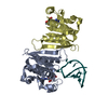
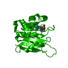
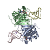
| Entry | Database: PDB / ID: 8h1a | ||||||
|---|---|---|---|---|---|---|---|
| Title | Crystal structure of MnmM from S. aureus in apo state (1.44 A) | ||||||
 Components Components | rRNA methylase YtqB | ||||||
 Keywords Keywords | TRANSFERASE / Methyltransferase / tRNA post-transcriptional modification / MnmC mnm5(s2)U | ||||||
| Function / homology |  Function and homology information Function and homology informationtRNA 5-(aminomethyl)-2-thiouridylate-methyltransferase / tRNA processing / methyltransferase activity / methylation Similarity search - Function | ||||||
| Biological species |  Staphylococcus aureus subsp. aureus NCTC 8325 (bacteria) Staphylococcus aureus subsp. aureus NCTC 8325 (bacteria) | ||||||
| Method |  X-RAY DIFFRACTION / SYNCHROTRON / MOLECULAR REPLACEMENT / Resolution: 1.44 Å X-RAY DIFFRACTION / SYNCHROTRON / MOLECULAR REPLACEMENT / Resolution: 1.44 Å | ||||||
 Authors Authors | Kim, J. / Cho, G. / Lee, J. | ||||||
| Funding support |  Korea, Republic Of, 1items Korea, Republic Of, 1items
| ||||||
 Citation Citation | Journal: Nucleic Acids Res. / Year: 2023 Title: Identification of a novel 5-aminomethyl-2-thiouridine methyltransferase in tRNA modification. Authors: Cho, G. / Lee, J. / Kim, J. | ||||||
| History |
|
- Structure visualization
| Structure viewer | Molecule: MolmilJmol/JSmol |
|---|
- Downloads & links
-Download
| PDBx/mmCIF format | 8h1a.cif.gz | 170.8 KB | Display | PDBx/mmCIF format |
|---|---|---|---|---|
| PDB format | pdb8h1a.ent.gz | 135.8 KB | Display | PDB format |
| PDBx/mmJSON format | 8h1a.json.gz | Tree view | PDBx/mmJSON format | |
| Others |  Other downloads Other downloads |
-Validation report
| Summary document | 8h1a_validation.pdf.gz | 432.2 KB | Display | wwPDB validaton report |
|---|---|---|---|---|
| Full document | 8h1a_full_validation.pdf.gz | 436.4 KB | Display | |
| Data in XML | 8h1a_validation.xml.gz | 18 KB | Display | |
| Data in CIF | 8h1a_validation.cif.gz | 26.4 KB | Display | |
| Arichive directory | https://data.pdbj.org/pub/pdb/validation_reports/h1/8h1aftp://data.pdbj.org/pub/pdb/validation_reports/h1/8h1a | HTTPS FTP |
-Related structure data

-Links
 PDBj
PDBj- Assembly
| Deposited unit | 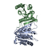
| ||||||||
|---|---|---|---|---|---|---|---|---|---|
| 1 |
| ||||||||
| Unit cell |
|
-Components
| #1: Protein | Mass: 21964.928 Da / Num. of mol.: 2 Source method: isolated from a genetically manipulated source Source: (gene. exp.) Staphylococcus aureus subsp. aureus NCTC 8325 (bacteria)Strain: NCTC 8325 / PS 47 / Gene: SAOUHSC_01878 / Plasmid: pLATE31 / Production host: #2: Water | ChemComp-HOH / |  Mass: 18.015 Da / Num. of mol.: 307 / Source method: isolated from a natural source / Formula: H2O Mass: 18.015 Da / Num. of mol.: 307 / Source method: isolated from a natural source / Formula: H2O |
|---|
-Experimental details
-Experiment
| Experiment | Method: X-RAY DIFFRACTION / Number of used crystals: 1 |
|---|
- Sample preparation
| Crystal | Density Matthews: 2.68 Å3/Da / Density % sol: 54.08 % |
|---|---|
| Crystal grow | Temperature: 293 K / Method: vapor diffusion, sitting drop Details: 0.2 M sodium acetate trihydrate, 0.1 M Tris-HCl, pH 8.5, 30% w/v PEG4000 |
-Data collection
| Diffraction | Mean temperature: 100 K / Ambient temp details: LN2 / Serial crystal experiment: N |
|---|---|
| Diffraction source | Source: SYNCHROTRON / Site: PAL/PLS / Beamline: 5C (4A) / Wavelength: 0.97965 Å |
| Detector | Type: DECTRIS EIGER X 9M / Detector: PIXEL / Date: Jun 29, 2022 |
| Radiation | Protocol: SINGLE WAVELENGTH / Monochromatic (M) / Laue (L): M / Scattering type: x-ray |
| Radiation wavelength | Wavelength: 0.97965 Å / Relative weight: 1 |
| Reflection | Resolution: 1.44→47.34 Å / Num. obs: 67371 / % possible obs: 95.5 % / Redundancy: 13.3 % / Biso Wilson estimate: 20.9 Å2 / CC1/2: 0.999 / Rmerge(I) obs: 0.073 / Rpim(I) all: 0.021 / Rrim(I) all: 0.076 / Net I/σ(I): 17.1 |
| Reflection shell | Resolution: 1.44→1.54 Å / Redundancy: 14.8 % / Rmerge(I) obs: 1.955 / Mean I/σ(I) obs: 1.5 / Num. unique obs: 3369 / CC1/2: 0.605 / Rpim(I) all: 0.525 / Rrim(I) all: 2.025 / % possible all: 66.5 |
- Processing
| Software |
| ||||||||||||||||||||||||||||||||||||||||||||||||||||||||||||||||||||||||||||||||||||||||||||||||||||||||||||||
|---|---|---|---|---|---|---|---|---|---|---|---|---|---|---|---|---|---|---|---|---|---|---|---|---|---|---|---|---|---|---|---|---|---|---|---|---|---|---|---|---|---|---|---|---|---|---|---|---|---|---|---|---|---|---|---|---|---|---|---|---|---|---|---|---|---|---|---|---|---|---|---|---|---|---|---|---|---|---|---|---|---|---|---|---|---|---|---|---|---|---|---|---|---|---|---|---|---|---|---|---|---|---|---|---|---|---|---|---|---|---|---|
| Refinement | Method to determine structure: MOLECULAR REPLACEMENT Starting model: AlphaFold model (UniProt: Q2FXG9) Resolution: 1.44→47.34 Å / Cor.coef. Fo:Fc: 0.974 / Cor.coef. Fo:Fc free: 0.963 / SU B: 3.075 / SU ML: 0.049 / Cross valid method: THROUGHOUT / ESU R: 0.072 / ESU R Free: 0.067 / Stereochemistry target values: MAXIMUM LIKELIHOOD Details: HYDROGENS HAVE BEEN ADDED IN THE RIDING POSITIONS U VALUES : REFINED INDIVIDUALLY
| ||||||||||||||||||||||||||||||||||||||||||||||||||||||||||||||||||||||||||||||||||||||||||||||||||||||||||||||
| Solvent computation | Ion probe radii: 0.9 Å / Shrinkage radii: 0.9 Å / VDW probe radii: 1.2 Å / Solvent model: MASK | ||||||||||||||||||||||||||||||||||||||||||||||||||||||||||||||||||||||||||||||||||||||||||||||||||||||||||||||
| Displacement parameters | Biso mean: 26.467 Å2
| ||||||||||||||||||||||||||||||||||||||||||||||||||||||||||||||||||||||||||||||||||||||||||||||||||||||||||||||
| Refinement step | Cycle: LAST / Resolution: 1.44→47.34 Å
| ||||||||||||||||||||||||||||||||||||||||||||||||||||||||||||||||||||||||||||||||||||||||||||||||||||||||||||||
| Refine LS restraints |
| ||||||||||||||||||||||||||||||||||||||||||||||||||||||||||||||||||||||||||||||||||||||||||||||||||||||||||||||
| LS refinement shell | Resolution: 1.44→1.476 Å
|
