 Movie
Movie Controller

[English] 日本語
 Yorodumi
- PDB-8gyg: Purification ,Crystallization and X-ray Diffraction analysis of a... -
+ Open data

- Basic information
| Entry | Database: PDB / ID: 8gyg | ||||||
|---|---|---|---|---|---|---|---|
| Title | Purification ,Crystallization and X-ray Diffraction analysis of a novel arysulfatase from Pseudoalteromonas atlantica T6c | ||||||
 Components Components | Arysulfatase | ||||||
 Keywords Keywords | HYDROLASE / Arysulfatase / Crystal | ||||||
| Function / homology | :  Function and homology information Function and homology information | ||||||
| Biological species |  Pseudoalteromonas atlantica T6c (bacteria) Pseudoalteromonas atlantica T6c (bacteria) | ||||||
| Method |  X-RAY DIFFRACTION / SYNCHROTRON / MOLECULAR REPLACEMENT / Resolution: 2 Å X-RAY DIFFRACTION / SYNCHROTRON / MOLECULAR REPLACEMENT / Resolution: 2 Å | ||||||
 Authors Authors | Dong, P.P. / Wu, Y.K. | ||||||
| Funding support |  China, 1items China, 1items
| ||||||
 Citation Citation | Journal: To Be Published Title: Purification ,Crystallization and X-ray Diffraction analysis of a novel arysulfatase from Pseudoalteromonas atlantica T6c Authors: Dong, P.P. / Wu, Y.K. | ||||||
| History |
|
- Structure visualization
| Structure viewer | Molecule: MolmilJmol/JSmol |
|---|
- Downloads & links
-Download
| PDBx/mmCIF format | 8gyg.cif.gz | 139.6 KB | Display | PDBx/mmCIF format |
|---|---|---|---|---|
| PDB format | pdb8gyg.ent.gz | 107.5 KB | Display | PDB format |
| PDBx/mmJSON format | 8gyg.json.gz | Tree view | PDBx/mmJSON format | |
| Others |  Other downloads Other downloads |
-Validation report
| Arichive directory | https://data.pdbj.org/pub/pdb/validation_reports/gy/8gygftp://data.pdbj.org/pub/pdb/validation_reports/gy/8gyg | HTTPS FTP |
|---|
-Related structure data
| Related structure data | 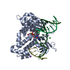 6nksS S: Starting model for refinement |
|---|---|
| Similar structure data |
-Links
 PDBj
PDBj- Assembly
| Deposited unit | 
| ||||||||
|---|---|---|---|---|---|---|---|---|---|
| 1 |
| ||||||||
| Unit cell |
|
-Components
| #1: Protein | Mass: 33342.402 Da / Num. of mol.: 2 Source method: isolated from a genetically manipulated source Source: (gene. exp.) Pseudoalteromonas atlantica T6c (bacteria)Production host: #2: Chemical | ChemComp-CA /   Mass: 40.078 Da / Num. of mol.: 4 / Source method: obtained synthetically / Formula: Ca Mass: 40.078 Da / Num. of mol.: 4 / Source method: obtained synthetically / Formula: Ca#3: Chemical | ChemComp-MN / |   Mass: 54.938 Da / Num. of mol.: 1 / Source method: obtained synthetically / Formula: Mn Mass: 54.938 Da / Num. of mol.: 1 / Source method: obtained synthetically / Formula: Mn#4: Water | ChemComp-HOH / |  Mass: 18.015 Da / Num. of mol.: 612 / Source method: isolated from a natural source / Formula: H2O Mass: 18.015 Da / Num. of mol.: 612 / Source method: isolated from a natural source / Formula: H2OHas ligand of interest | N | Has protein modification | N | |
|---|
-Experimental details
-Experiment
| Experiment | Method: X-RAY DIFFRACTION / Number of used crystals: 1 |
|---|
- Sample preparation
| Crystal | Density Matthews: 2.27 Å3/Da / Density % sol: 45.89 % / Description: strip |
|---|---|
| Crystal grow | Temperature: 297 K / Method: vapor diffusion, sitting drop / pH: 8 / Details: 2o%PEG 3350, 0.2M Sodium Thiocyamate |
-Data collection
| Diffraction | Mean temperature: 100 K / Serial crystal experiment: N |
|---|---|
| Diffraction source | Source: SYNCHROTRON / Site: SSRF / Beamline: BL18U1 / Wavelength: 0.97915 Å |
| Detector | Type: DECTRIS PILATUS3 S 6M / Detector: PIXEL / Date: Jan 6, 2022 |
| Radiation | Protocol: SINGLE WAVELENGTH / Monochromatic (M) / Laue (L): M / Scattering type: x-ray |
| Radiation wavelength | Wavelength: 0.97915 Å / Relative weight: 1 |
| Reflection | Resolution: 1.55→38.61 Å / Num. obs: 85833 / % possible obs: 99.7 % / Redundancy: 6.1 % / CC1/2: 0.995 / Rmerge(I) obs: 0.117 / Net I/σ(I): 8.3 |
| Reflection shell | Resolution: 1.55→1.59 Å / Rmerge(I) obs: 1.077 / Num. unique obs: 6332 / CC1/2: 0.528 |
- Processing
| Software |
| ||||||||||||||||||||||||||||||||||||||||||||||||||||||||||||||||||||||||||||||||||||||||||
|---|---|---|---|---|---|---|---|---|---|---|---|---|---|---|---|---|---|---|---|---|---|---|---|---|---|---|---|---|---|---|---|---|---|---|---|---|---|---|---|---|---|---|---|---|---|---|---|---|---|---|---|---|---|---|---|---|---|---|---|---|---|---|---|---|---|---|---|---|---|---|---|---|---|---|---|---|---|---|---|---|---|---|---|---|---|---|---|---|---|---|---|
| Refinement | Method to determine structure: MOLECULAR REPLACEMENT Starting model: 6nks Resolution: 2→29.585 Å / SU ML: 0.24 / Cross valid method: THROUGHOUT / σ(F): 1.38 / Phase error: 25.73 / Stereochemistry target values: ML
| ||||||||||||||||||||||||||||||||||||||||||||||||||||||||||||||||||||||||||||||||||||||||||
| Solvent computation | Shrinkage radii: 0.9 Å / VDW probe radii: 1.11 Å / Solvent model: FLAT BULK SOLVENT MODEL | ||||||||||||||||||||||||||||||||||||||||||||||||||||||||||||||||||||||||||||||||||||||||||
| Displacement parameters | Biso max: 66.52 Å2 / Biso mean: 23.9035 Å2 / Biso min: 7.36 Å2 | ||||||||||||||||||||||||||||||||||||||||||||||||||||||||||||||||||||||||||||||||||||||||||
| Refinement step | Cycle: final / Resolution: 2→29.585 Å
| ||||||||||||||||||||||||||||||||||||||||||||||||||||||||||||||||||||||||||||||||||||||||||
| LS refinement shell | Refine-ID: X-RAY DIFFRACTION / Rfactor Rfree error: 0
|
