 Movie
Movie Controller


+ Open data

- Basic information
| Entry | Database: PDB / ID: 8gx9 | ||||||
|---|---|---|---|---|---|---|---|
| Title | Crystal structure of SARS-CoV-2 RBD with P2C-1F11 and P2B-1G5 | ||||||
 Components Components |
| ||||||
 Keywords Keywords | VIRAL PROTEIN/IMMUNE SYSTEM / spike / receptor binding domain / antibody / viral protein / VIRAL PROTEIN-IMMUNE SYSTEM complex | ||||||
| Function / homology |  Function and homology information Function and homology informationMaturation of spike protein / viral translation / Translation of Structural Proteins / Virion Assembly and Release / host cell surface / host extracellular space / suppression by virus of host tetherin activity / Induction of Cell-Cell Fusion / structural constituent of virion / entry receptor-mediated virion attachment to host cell ...Maturation of spike protein / viral translation / Translation of Structural Proteins / Virion Assembly and Release / host cell surface / host extracellular space / suppression by virus of host tetherin activity / Induction of Cell-Cell Fusion / structural constituent of virion / entry receptor-mediated virion attachment to host cell / host cell endoplasmic reticulum-Golgi intermediate compartment membrane / receptor-mediated endocytosis of virus by host cell / membrane fusion / Attachment and Entry / positive regulation of viral entry into host cell / receptor-mediated virion attachment to host cell / receptor ligand activity / host cell surface receptor binding / fusion of virus membrane with host plasma membrane / fusion of virus membrane with host endosome membrane / viral envelope / virion attachment to host cell / SARS-CoV-2 activates/modulates innate and adaptive immune responses / host cell plasma membrane / virion membrane / identical protein binding / membrane / plasma membrane Similarity search - Function | ||||||
| Biological species |   Severe acute respiratory syndrome coronavirus 2 Severe acute respiratory syndrome coronavirus 2 Homo sapiens (human) Homo sapiens (human) | ||||||
| Method |  X-RAY DIFFRACTION / SYNCHROTRON / MOLECULAR REPLACEMENT / Resolution: 4.01 Å X-RAY DIFFRACTION / SYNCHROTRON / MOLECULAR REPLACEMENT / Resolution: 4.01 Å | ||||||
 Authors Authors | Wang, X. / Zhang, L. / Ge, J. | ||||||
| Funding support |  China, 1items China, 1items
| ||||||
 Citation Citation | Journal: To be published Title: Crystal structure of SARS-CoV-2 antibody P2C-1F11 and RBD Authors: Wang, X. / Zhang, L. / Ge, J. | ||||||
| History |
|
- Structure visualization
| Structure viewer | Molecule: MolmilJmol/JSmol |
|---|
- Downloads & links
-Download
| PDBx/mmCIF format | 8gx9.cif.gz | 400.3 KB | Display | PDBx/mmCIF format |
|---|---|---|---|---|
| PDB format | pdb8gx9.ent.gz | 321.9 KB | Display | PDB format |
| PDBx/mmJSON format | 8gx9.json.gz | Tree view | PDBx/mmJSON format | |
| Others |  Other downloads Other downloads |
-Validation report
| Summary document | 8gx9_validation.pdf.gz | 523.2 KB | Display | wwPDB validaton report |
|---|---|---|---|---|
| Full document | 8gx9_full_validation.pdf.gz | 550.7 KB | Display | |
| Data in XML | 8gx9_validation.xml.gz | 71.3 KB | Display | |
| Data in CIF | 8gx9_validation.cif.gz | 96.2 KB | Display | |
| Arichive directory | https://data.pdbj.org/pub/pdb/validation_reports/gx/8gx9ftp://data.pdbj.org/pub/pdb/validation_reports/gx/8gx9 | HTTPS FTP |
-Related structure data
| Related structure data | 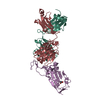 7cdiS S: Starting model for refinement |
|---|---|
| Similar structure data |
-Links
 PDBj
PDBj


- Assembly
| Deposited unit | 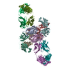
| ||||||||
|---|---|---|---|---|---|---|---|---|---|
| 1 |
| ||||||||
| Unit cell |
|
-Components
-Antibody , 3 types, 6 molecules FLHIMN
| #2: Antibody | Mass: 23274.766 Da / Num. of mol.: 2 Source method: isolated from a genetically manipulated source Source: (gene. exp.) Homo sapiens (human) / Production host: Homo sapiens (human)#3: Antibody | Mass: 22754.477 Da / Num. of mol.: 2 Source method: isolated from a genetically manipulated source Source: (gene. exp.) Homo sapiens (human) / Production host: Homo sapiens (human)#4: Antibody | Mass: 22778.186 Da / Num. of mol.: 2 Source method: isolated from a genetically manipulated source Source: (gene. exp.) Homo sapiens (human) / Production host: Homo sapiens (human) |
|---|
-Protein / Heavy chain of ... / Sugars , 3 types, 6 molecules AEOP
| #1: Protein | Mass: 23771.750 Da / Num. of mol.: 2 Source method: isolated from a genetically manipulated source Source: (gene. exp.) Severe acute respiratory syndrome coronavirus 2Gene: S, 2 / Production host:   Spodoptera frugiperda (fall armyworm) / References: UniProt: P0DTC2 Spodoptera frugiperda (fall armyworm) / References: UniProt: P0DTC2#5: Protein | Mass: 22912.660 Da / Num. of mol.: 2 Source method: isolated from a genetically manipulated source Source: (gene. exp.) Homo sapiens (human) / Production host: Homo sapiens (human)#6: Sugar |  Type: D-saccharide, beta linking / Mass: 221.208 Da / Num. of mol.: 2 / Source method: obtained synthetically / Formula: C8H15NO6 Type: D-saccharide, beta linking / Mass: 221.208 Da / Num. of mol.: 2 / Source method: obtained synthetically / Formula: C8H15NO6 |
|---|
-Details
| Has ligand of interest | N |
|---|
-Experimental details
-Experiment
| Experiment | Method: X-RAY DIFFRACTION / Number of used crystals: 1 |
|---|
- Sample preparation
| Crystal | Density Matthews: 2.97 Å3/Da / Density % sol: 58.56 % |
|---|---|
| Crystal grow | Temperature: 291.15 K / Method: vapor diffusion, sitting drop Details: 0.2M sodium chloride, 0.1M MES, pH 6.0, 20% PEG 2000 MME |
-Data collection
| Diffraction | Mean temperature: 100 K / Serial crystal experiment: N |
|---|---|
| Diffraction source | Source: SYNCHROTRON / Site: SSRF / Beamline: BL17U1 / Wavelength: 0.9769 Å |
| Detector | Type: DECTRIS EIGER X 16M / Detector: PIXEL / Date: Oct 22, 2020 |
| Radiation | Protocol: SINGLE WAVELENGTH / Monochromatic (M) / Laue (L): M / Scattering type: x-ray |
| Radiation wavelength | Wavelength: 0.9769 Å / Relative weight: 1 |
| Reflection | Resolution: 4.01→49.08 Å / Num. obs: 23031 / % possible obs: 97.27 % / Redundancy: 6.3 % / Rmerge(I) obs: 0.29 / Net I/σ(I): 4.8 |
| Reflection shell | Resolution: 4.01→4.153 Å / Num. unique obs: 2097 / CC1/2: 0.505 |
- Processing
| Software |
| ||||||||||||||||||||||||||||||||||||||||||||||||||||||||||||||||||||||
|---|---|---|---|---|---|---|---|---|---|---|---|---|---|---|---|---|---|---|---|---|---|---|---|---|---|---|---|---|---|---|---|---|---|---|---|---|---|---|---|---|---|---|---|---|---|---|---|---|---|---|---|---|---|---|---|---|---|---|---|---|---|---|---|---|---|---|---|---|---|---|---|
| Refinement | Method to determine structure: MOLECULAR REPLACEMENT Starting model: 7CDI Resolution: 4.01→49.08 Å / SU ML: 0.54 / Cross valid method: THROUGHOUT / σ(F): 1.33 / Phase error: 34.3 / Stereochemistry target values: ML
| ||||||||||||||||||||||||||||||||||||||||||||||||||||||||||||||||||||||
| Solvent computation | Shrinkage radii: 0.9 Å / VDW probe radii: 1.11 Å / Solvent model: FLAT BULK SOLVENT MODEL | ||||||||||||||||||||||||||||||||||||||||||||||||||||||||||||||||||||||
| Displacement parameters | Biso max: 215.21 Å2 / Biso mean: 139.9726 Å2 / Biso min: 30 Å2 | ||||||||||||||||||||||||||||||||||||||||||||||||||||||||||||||||||||||
| Refinement step | Cycle: final / Resolution: 4.01→49.08 Å
| ||||||||||||||||||||||||||||||||||||||||||||||||||||||||||||||||||||||
| LS refinement shell | Refine-ID: X-RAY DIFFRACTION / Rfactor Rfree error: 0 / Total num. of bins used: 9
|
