 ムービー
ムービー コントローラー


+ データを開く

- 基本情報
基本情報
| 登録情報 | データベース: PDB / ID: 8gwa | ||||||||||||||||||||||||||||||||||||||||||||||||||||||||||||
|---|---|---|---|---|---|---|---|---|---|---|---|---|---|---|---|---|---|---|---|---|---|---|---|---|---|---|---|---|---|---|---|---|---|---|---|---|---|---|---|---|---|---|---|---|---|---|---|---|---|---|---|---|---|---|---|---|---|---|---|---|---|
| タイトル | Structure of the intact photosynthetic light-harvesting antenna-reaction center complex from a green sulfur bacterium | ||||||||||||||||||||||||||||||||||||||||||||||||||||||||||||
 要素 要素 |
| ||||||||||||||||||||||||||||||||||||||||||||||||||||||||||||
 キーワード キーワード | PHOTOSYNTHESIS / Green sulfur bacterium / Reaction center / FMO protein / Energy transfer | ||||||||||||||||||||||||||||||||||||||||||||||||||||||||||||
| 機能・相同性 |  機能・相同性情報 機能・相同性情報thylakoid / bacteriochlorophyll binding / iron-sulfur cluster binding / photosynthesis / electron transfer activity / heme binding / membrane / metal ion binding / plasma membrane 類似検索 - 分子機能 | ||||||||||||||||||||||||||||||||||||||||||||||||||||||||||||
| 生物種 |  Chlorobaculum tepidum TLS (バクテリア) Chlorobaculum tepidum TLS (バクテリア) | ||||||||||||||||||||||||||||||||||||||||||||||||||||||||||||
| 手法 | 電子顕微鏡法 / 単粒子再構成法 / クライオ電子顕微鏡法 / 解像度: 2.9 Å | ||||||||||||||||||||||||||||||||||||||||||||||||||||||||||||
 データ登録者 データ登録者 | Chen, J.H. / Zhang, X. | ||||||||||||||||||||||||||||||||||||||||||||||||||||||||||||
| 資金援助 |  中国, 1件 中国, 1件
| ||||||||||||||||||||||||||||||||||||||||||||||||||||||||||||
 引用 引用 | ジャーナル: J Integr Plant Biol / 年: 2023 タイトル: Cryo-electron microscopy structure of the intact photosynthetic light-harvesting antenna-reaction center complex from a green sulfur bacterium. 著者: Jing-Hua Chen / Weiwei Wang / Chen Wang / Tingyun Kuang / Jian-Ren Shen / Xing Zhang /  要旨: The photosynthetic reaction center complex (RCC) of green sulfur bacteria (GSB) consists of the membrane-imbedded RC core and the peripheric energy transmitting proteins called Fenna-Matthews-Olson ...The photosynthetic reaction center complex (RCC) of green sulfur bacteria (GSB) consists of the membrane-imbedded RC core and the peripheric energy transmitting proteins called Fenna-Matthews-Olson (FMO). Functionally, FMO transfers the absorbed energy from a huge peripheral light-harvesting antenna named chlorosome to the RC core where charge separation occurs. In vivo, one RC was found to bind two FMOs, however, the intact structure of RCC as well as the energy transfer mechanism within RCC remain to be clarified. Here we report a structure of intact RCC which contains a RC core and two FMO trimers from a thermophilic green sulfur bacterium Chlorobaculum tepidum at 2.9 Å resolution by cryo-electron microscopy. The second FMO trimer is attached at the cytoplasmic side asymmetrically relative to the first FMO trimer reported previously. We also observed two new subunits (PscE and PscF) and the N-terminal transmembrane domain of a cytochrome-containing subunit (PscC) in the structure. These two novel subunits possibly function to facilitate the binding of FMOs to RC core and to stabilize the whole complex. A new bacteriochlorophyll (numbered as 816) was identified at the interspace between PscF and PscA-1, causing an asymmetrical energy transfer from the two FMO trimers to RC core. Based on the structure, we propose an energy transfer network within this photosynthetic apparatus. | ||||||||||||||||||||||||||||||||||||||||||||||||||||||||||||
| 履歴 |
|
- 構造の表示
構造の表示
| 構造ビューア | 分子: MolmilJmol/JSmol |
|---|
- ダウンロードとリンク
-ダウンロード
| PDBx/mmCIF形式 | 8gwa.cif.gz | 831.8 KB | 表示 | PDBx/mmCIF形式 |
|---|---|---|---|---|
| PDB形式 | pdb8gwa.ent.gz | 表示 | PDB形式 | |
| PDBx/mmJSON形式 | 8gwa.json.gz | ツリー表示 | PDBx/mmJSON形式 | |
| その他 |  その他のダウンロード その他のダウンロード |
-検証レポート
| アーカイブディレクトリ | https://data.pdbj.org/pub/pdb/validation_reports/gw/8gwaftp://data.pdbj.org/pub/pdb/validation_reports/gw/8gwa | HTTPS FTP |
|---|
-関連構造データ
-リンク
 PDBj
PDBj


- 集合体
集合体
| 登録構造単位 | 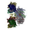
|
|---|---|
| 1 |
|
-要素
-Bacteriochlorophyll ... , 1種, 6分子 132465
| #1: タンパク質 | 分子量: 40343.430 Da / 分子数: 6 / 由来タイプ: 天然 由来: (天然) Chlorobaculum tepidum TLS (バクテリア)株: ATCC 49652 / DSM 12025 / NBRC 103806 / TLS / 参照: UniProt: Q46393 |
|---|
-タンパク質 , 4種, 5分子 DEFCc
| #2: タンパク質 | 分子量: 16633.195 Da / 分子数: 1 / 由来タイプ: 天然 由来: (天然) Chlorobaculum tepidum TLS (バクテリア)株: ATCC 49652 / DSM 12025 / NBRC 103806 / TLS / 参照: UniProt: Q8KEP5 |
|---|---|
| #5: タンパク質 | 分子量: 4954.098 Da / 分子数: 1 / 由来タイプ: 天然 由来: (天然) Chlorobaculum tepidum TLS (バクテリア) |
| #6: タンパク質 | 分子量: 6227.533 Da / 分子数: 1 / 由来タイプ: 天然 由来: (天然) Chlorobaculum tepidum TLS (バクテリア)株: ATCC 49652 / DSM 12025 / NBRC 103806 / TLS / 参照: UniProt: Q8KG87 |
| #7: タンパク質 | 分子量: 22741.779 Da / 分子数: 2 / 由来タイプ: 天然 由来: (天然) Chlorobaculum tepidum TLS (バクテリア)株: ATCC 49652 / DSM 12025 / NBRC 103806 / TLS / 参照: UniProt: O07091 |
-Photosystem P840 reaction ... , 2種, 3分子 BAa
| #3: タンパク質 | 分子量: 23383.096 Da / 分子数: 1 / 由来タイプ: 天然 由来: (天然) Chlorobaculum tepidum TLS (バクテリア)株: ATCC 49652 / DSM 12025 / NBRC 103806 / TLS / 参照: UniProt: Q8KAY1 |
|---|---|
| #4: タンパク質 | 分子量: 81784.641 Da / 分子数: 2 / 由来タイプ: 天然 由来: (天然) Chlorobaculum tepidum TLS (バクテリア)参照: UniProt: Q8KAY0 |
-非ポリマー , 10種, 103分子 


| #8: 化合物 | ChemComp-BCL /  分子量: 911.504 Da / 分子数: 73 / 由来タイプ: 合成 / 式: C55H74MgN4O6 分子量: 911.504 Da / 分子数: 73 / 由来タイプ: 合成 / 式: C55H74MgN4O6#9: 化合物 | ChemComp-CDL /  分子量: 1464.043 Da / 分子数: 4 / 由来タイプ: 合成 / 式: C81H156O17P2 / タイプ: SUBJECT OF INVESTIGATION / コメント: リン脂質*YM 分子量: 1464.043 Da / 分子数: 4 / 由来タイプ: 合成 / 式: C81H156O17P2 / タイプ: SUBJECT OF INVESTIGATION / コメント: リン脂質*YM#10: 化合物 |  分子量: 351.640 Da / 分子数: 3 / 由来タイプ: 合成 / 式: Fe4S4 分子量: 351.640 Da / 分子数: 3 / 由来タイプ: 合成 / 式: Fe4S4#11: 化合物 |  分子量: 911.504 Da / 分子数: 2 / 由来タイプ: 合成 / 式: C55H74MgN4O6 / タイプ: SUBJECT OF INVESTIGATION 分子量: 911.504 Da / 分子数: 2 / 由来タイプ: 合成 / 式: C55H74MgN4O6 / タイプ: SUBJECT OF INVESTIGATION#12: 化合物 | ChemComp-G2O /  分子量: 891.473 Da / 分子数: 4 / 由来タイプ: 合成 / 式: C55H70MgN4O5 / タイプ: SUBJECT OF INVESTIGATION 分子量: 891.473 Da / 分子数: 4 / 由来タイプ: 合成 / 式: C55H70MgN4O5 / タイプ: SUBJECT OF INVESTIGATION#13: 化合物 | ChemComp-F39 / [(  分子量: 895.299 Da / 分子数: 4 / 由来タイプ: 合成 / 式: C58H86O7 分子量: 895.299 Da / 分子数: 4 / 由来タイプ: 合成 / 式: C58H86O7#14: 化合物 | ChemComp-LHG /  分子量: 722.970 Da / 分子数: 7 / 由来タイプ: 合成 / 式: C38H75O10P / コメント: リン脂質*YM 分子量: 722.970 Da / 分子数: 7 / 由来タイプ: 合成 / 式: C38H75O10P / コメント: リン脂質*YM#15: 化合物 |  分子量: 787.158 Da / 分子数: 2 / 由来タイプ: 合成 / 式: C45H86O10 分子量: 787.158 Da / 分子数: 2 / 由来タイプ: 合成 / 式: C45H86O10#16: 化合物 |  分子量: 40.078 Da / 分子数: 2 / 由来タイプ: 合成 / 式: Ca 分子量: 40.078 Da / 分子数: 2 / 由来タイプ: 合成 / 式: Ca#17: 化合物 |  分子量: 532.841 Da / 分子数: 2 / 由来タイプ: 合成 / 式: C40H52 分子量: 532.841 Da / 分子数: 2 / 由来タイプ: 合成 / 式: C40H52 |
|---|
-詳細
| 研究の焦点であるリガンドがあるか | Y |
|---|---|
| Has protein modification | N |
-実験情報
-実験
| 実験 | 手法: 電子顕微鏡法 |
|---|---|
| EM実験 | 試料の集合状態: PARTICLE / 3次元再構成法: 単粒子再構成法 |
- 試料調製
試料調製
| 構成要素 | 名称: GsbRC-2FMO complex / タイプ: COMPLEX / Entity ID: #1-#7 / 由来: NATURAL |
|---|---|
| 分子量 | 実験値: NO |
| 由来(天然) | 生物種: Chlorobaculum tepidum TLS (バクテリア) |
| 緩衝液 | pH: 8 |
| 試料 | 包埋: NO / シャドウイング: NO / 染色: NO / 凍結: YES |
| 急速凍結 | 装置: FEI VITROBOT MARK IV / 凍結剤: ETHANE / 湿度: 100 % / 凍結前の試料温度: 277 K |
- 電子顕微鏡撮影
| 実験機器 |  モデル: Titan Krios / 画像提供: FEI Company |
|---|---|
| 顕微鏡 | モデル: FEI TITAN KRIOS |
| 電子銃 | 電子線源:  FIELD EMISSION GUN / 加速電圧: 300 kV / 照射モード: FLOOD BEAM FIELD EMISSION GUN / 加速電圧: 300 kV / 照射モード: FLOOD BEAM |
| 電子レンズ | モード: BRIGHT FIELD / 倍率(公称値): 130000 X / 倍率(補正後): 152200 X / 最大 デフォーカス(公称値): 1500 nm / 最小 デフォーカス(公称値): 800 nm / アライメント法: COMA FREE |
| 試料ホルダ | 凍結剤: NITROGEN 試料ホルダーモデル: FEI TITAN KRIOS AUTOGRID HOLDER 最高温度: 90 K / 最低温度: 80 K |
| 撮影 | 平均露光時間: 8 sec. / 電子線照射量: 60 e/Å2 フィルム・検出器のモデル: FEI FALCON IV (4k x 4k) 撮影したグリッド数: 1 / 実像数: 2297 |
| 電子光学装置 | エネルギーフィルター名称: TFS Selectris / 色収差補正装置: 2.7mm / エネルギーフィルタースリット幅: 10 eV / 球面収差補正装置: 2.7mm |
- 解析
解析
| ソフトウェア | 名称: PHENIX / バージョン: 1.19.2_4158: / 分類: 精密化 | ||||||||||||||||||||||||||||||||
|---|---|---|---|---|---|---|---|---|---|---|---|---|---|---|---|---|---|---|---|---|---|---|---|---|---|---|---|---|---|---|---|---|---|
| EMソフトウェア |
| ||||||||||||||||||||||||||||||||
| CTF補正 | タイプ: PHASE FLIPPING AND AMPLITUDE CORRECTION | ||||||||||||||||||||||||||||||||
| 粒子像の選択 | 選択した粒子像数: 1961370 | ||||||||||||||||||||||||||||||||
| 対称性 | 点対称性: C1 (非対称) | ||||||||||||||||||||||||||||||||
| 3次元再構成 | 解像度: 2.9 Å / 解像度の算出法: FSC 0.143 CUT-OFF / 粒子像の数: 227038 / クラス平均像の数: 1 / 対称性のタイプ: POINT | ||||||||||||||||||||||||||||||||
| 拘束条件 |
|
