 Movie
Movie Controller


+ Open data

- Basic information
| Entry | Database: PDB / ID: 8gw6 | |||||||||||||||
|---|---|---|---|---|---|---|---|---|---|---|---|---|---|---|---|---|
| Title | AtSLAC1 6D mutant in closed state | |||||||||||||||
 Components Components | Guard cell S-type anion channel SLAC1,Green fluorescent protein (Fragment) | |||||||||||||||
 Keywords Keywords | MEMBRANE PROTEIN / Stomatal closure / anion channel / phosphorylation-dependent activation | |||||||||||||||
| Function / homology |  Function and homology information Function and homology informationresponse to humidity / stomatal closure / regulation of stomatal opening / inorganic anion transport / voltage-gated monoatomic anion channel activity / regulation of stomatal closure / stomatal movement / response to ozone / response to carbon dioxide / intracellular monoatomic ion homeostasis ...response to humidity / stomatal closure / regulation of stomatal opening / inorganic anion transport / voltage-gated monoatomic anion channel activity / regulation of stomatal closure / stomatal movement / response to ozone / response to carbon dioxide / intracellular monoatomic ion homeostasis / monoatomic anion transmembrane transporter activity / multicellular organismal-level water homeostasis / : / response to abscisic acid / abscisic acid-activated signaling pathway / monoatomic anion transport / response to light stimulus / bioluminescence / generation of precursor metabolites and energy / protein phosphatase binding / protein kinase binding / membrane / plasma membrane Similarity search - Function | |||||||||||||||
| Biological species |  | |||||||||||||||
| Method | ELECTRON MICROSCOPY / single particle reconstruction / cryo EM / Resolution: 3.3 Å | |||||||||||||||
 Authors Authors | Lee, Y. / Lee, S. | |||||||||||||||
| Funding support |  Korea, Republic Of, 4items Korea, Republic Of, 4items
| |||||||||||||||
 Citation Citation | Journal: Nat Commun / Year: 2023 Title: Cryo-EM structures of the plant anion channel SLAC1 from Arabidopsis thaliana suggest a combined activation model. Authors: Yeongmok Lee / Hyeon Seong Jeong / Seoyeon Jung / Junmo Hwang / Chi Truc Han Le / Sung-Hoon Jun / Eun Jo Du / KyeongJin Kang / Beom-Gi Kim / Hyun-Ho Lim / Sangho Lee / Abstract: The anion channel SLAC1 functions as a crucial effector in the ABA signaling, leading to stomata closure. SLAC1 is activated by phosphorylation in its intracellular domains. Both a binding-activation ...The anion channel SLAC1 functions as a crucial effector in the ABA signaling, leading to stomata closure. SLAC1 is activated by phosphorylation in its intracellular domains. Both a binding-activation model and an inhibition-release model for activation have been proposed based on only the closed structures of SLAC1, rendering the structure-based activation mechanism controversial. Here we report cryo-EM structures of Arabidopsis SLAC1 WT and its phosphomimetic mutants in open and closed states. Comparison of the open structure with the closed ones reveals the structural basis for opening of the conductance pore. Multiple phosphorylation of an intracellular domain (ICD) causes dissociation of ICD from the transmembrane domain. A conserved, positively-charged sequence motif in the intracellular loop 2 (ICL2) seems to be capable of sensing of the negatively charged phosphorylated ICD. Interactions between ICL2 and ICD drive drastic conformational changes, thereby widening the pore. From our results we propose that SLAC1 operates by a mechanism combining the binding-activation and inhibition-release models. | |||||||||||||||
| History |
|
- Structure visualization
| Structure viewer | Molecule: MolmilJmol/JSmol |
|---|
- Downloads & links
-Download
| PDBx/mmCIF format | 8gw6.cif.gz | 229.8 KB | Display | PDBx/mmCIF format |
|---|---|---|---|---|
| PDB format | pdb8gw6.ent.gz | 171.1 KB | Display | PDB format |
| PDBx/mmJSON format | 8gw6.json.gz | Tree view | PDBx/mmJSON format | |
| Others |  Other downloads Other downloads |
-Validation report
| Arichive directory | https://data.pdbj.org/pub/pdb/validation_reports/gw/8gw6ftp://data.pdbj.org/pub/pdb/validation_reports/gw/8gw6 | HTTPS FTP |
|---|
-Related structure data
| Related structure data | 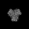 34303MC 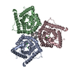 8gw7C 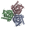 8j0jC 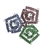 8j1eC M: map data used to model this data C: citing same article ( |
|---|---|
| Similar structure data |
-Links
 PDBj
PDBj

- Assembly
| Deposited unit | 
| ||||||||||||||||||||||||||||||||||||||||||||||||||||||||||||||||||||||||||||||||||||||||||||||
|---|---|---|---|---|---|---|---|---|---|---|---|---|---|---|---|---|---|---|---|---|---|---|---|---|---|---|---|---|---|---|---|---|---|---|---|---|---|---|---|---|---|---|---|---|---|---|---|---|---|---|---|---|---|---|---|---|---|---|---|---|---|---|---|---|---|---|---|---|---|---|---|---|---|---|---|---|---|---|---|---|---|---|---|---|---|---|---|---|---|---|---|---|---|---|---|
| 1 |
| ||||||||||||||||||||||||||||||||||||||||||||||||||||||||||||||||||||||||||||||||||||||||||||||
| Noncrystallographic symmetry (NCS) | NCS domain:
NCS domain segments:
NCS oper:
|
-Components
| #1: Protein | Mass: 94016.984 Da / Num. of mol.: 3 / Mutation: T62D,S65D,S107D,S124D,S146D,T152D Source method: isolated from a genetically manipulated source Source: (gene. exp.) Gene: SLAC1, CDI3, OZS1, RCD3, At1g12480, F5O11.23, T12C24.3, gfp Plasmid: pACEBac2 / Cell line (production host): Sf9 / Production host:   Spodoptera frugiperda (fall armyworm) / References: UniProt: Q9LD83, UniProt: A0A059PIQ0 Spodoptera frugiperda (fall armyworm) / References: UniProt: Q9LD83, UniProt: A0A059PIQ0#2: Chemical |   Mass: 35.453 Da / Num. of mol.: 3 / Source method: obtained synthetically / Formula: Cl Mass: 35.453 Da / Num. of mol.: 3 / Source method: obtained synthetically / Formula: Cl#3: Chemical |   Mass: 486.726 Da / Num. of mol.: 3 / Source method: obtained synthetically / Formula: C31H50O4 Mass: 486.726 Da / Num. of mol.: 3 / Source method: obtained synthetically / Formula: C31H50O4Has ligand of interest | N | |
|---|
-Experimental details
-Experiment
| Experiment | Method: ELECTRON MICROSCOPY |
|---|---|
| EM experiment | Aggregation state: PARTICLE / 3D reconstruction method: single particle reconstruction |
- Sample preparation
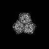
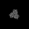
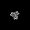
| Component | Name: Trimer of closed state AtSLAC1 6D mutant with sfGFP tag in GDN and CHS micelle Type: COMPLEX / Entity ID: #1 / Source: RECOMBINANT | ||||||||||||||||||||||||||||||
|---|---|---|---|---|---|---|---|---|---|---|---|---|---|---|---|---|---|---|---|---|---|---|---|---|---|---|---|---|---|---|---|
| Molecular weight | Value: 0.28 MDa / Experimental value: NO | ||||||||||||||||||||||||||||||
| Source (natural) | Organism: | ||||||||||||||||||||||||||||||
| Source (recombinant) | Organism: Spodoptera frugiperda (fall armyworm) / Strain: Sf9 | ||||||||||||||||||||||||||||||
| Buffer solution | pH: 7 | ||||||||||||||||||||||||||||||
| Buffer component |
| ||||||||||||||||||||||||||||||
| Specimen | Conc.: 3.3 mg/ml / Embedding applied: NO / Shadowing applied: NO / Staining applied: NO / Vitrification applied: YES | ||||||||||||||||||||||||||||||
| Specimen support | Grid material: GOLD / Grid mesh size: 300 divisions/in. / Grid type: Quantifoil R1.2/1.3 | ||||||||||||||||||||||||||||||
| Vitrification | Instrument: FEI VITROBOT MARK III / Cryogen name: ETHANE / Humidity: 100 % / Chamber temperature: 277 K |
- Electron microscopy imaging
| Experimental equipment |  Model: Titan Krios / Image courtesy: FEI Company |
|---|---|
| Microscopy | Model: TFS KRIOS |
| Electron gun | Electron source:  FIELD EMISSION GUN / Accelerating voltage: 300 kV / Illumination mode: FLOOD BEAM FIELD EMISSION GUN / Accelerating voltage: 300 kV / Illumination mode: FLOOD BEAM |
| Electron lens | Mode: BRIGHT FIELD / Nominal defocus max: 1900 nm / Nominal defocus min: 700 nm / Cs: 2.7 mm / C2 aperture diameter: 70 µm |
| Specimen holder | Cryogen: NITROGEN |
| Image recording | Electron dose: 50 e/Å2 / Film or detector model: GATAN K3 BIOQUANTUM (6k x 4k) |
| EM imaging optics | Energyfilter name: GIF Bioquantum / Energyfilter slit width: 14 eV |
- Processing
| Software |
| ||||||||||||||||||||||||
|---|---|---|---|---|---|---|---|---|---|---|---|---|---|---|---|---|---|---|---|---|---|---|---|---|---|
| EM software |
| ||||||||||||||||||||||||
| CTF correction | Type: NONE | ||||||||||||||||||||||||
| Symmetry | Point symmetry: C3 (3 fold cyclic) | ||||||||||||||||||||||||
| 3D reconstruction | Resolution: 3.3 Å / Resolution method: FSC 0.143 CUT-OFF / Num. of particles: 69390 / Algorithm: FOURIER SPACE / Symmetry type: POINT | ||||||||||||||||||||||||
| Refinement | Cross valid method: NONE | ||||||||||||||||||||||||
| Refine LS restraints |
| ||||||||||||||||||||||||
| Refine LS restraints NCS |
|
