National Natural Science Foundation of China (NSFC)
52021006
China
Citation
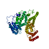
Journal: Nat Methods / Year: 2023 Title: Uniform thin ice on ultraflat graphene for high-resolution cryo-EM. Authors: Liming Zheng / Nan Liu / Xiaoyin Gao / Wenqing Zhu / Kun Liu / Cang Wu / Rui Yan / Jincan Zhang / Xin Gao / Yating Yao / Bing Deng / Jie Xu / Ye Lu / Zhongmin Liu / Mengsen Li / Xiaoding Wei ...Authors: Liming Zheng / Nan Liu / Xiaoyin Gao / Wenqing Zhu / Kun Liu / Cang Wu / Rui Yan / Jincan Zhang / Xin Gao / Yating Yao / Bing Deng / Jie Xu / Ye Lu / Zhongmin Liu / Mengsen Li / Xiaoding Wei / Hong-Wei Wang / Hailin Peng / Abstract: Cryo-electron microscopy (cryo-EM) visualizes the atomic structure of macromolecules that are embedded in vitrified thin ice at their close-to-native state. However, the homogeneity of ice thickness, ...Cryo-electron microscopy (cryo-EM) visualizes the atomic structure of macromolecules that are embedded in vitrified thin ice at their close-to-native state. However, the homogeneity of ice thickness, a key factor to ensure high image quality, is poorly controlled during specimen preparation and has become one of the main challenges for high-resolution cryo-EM. Here we found that the uniformity of thin ice relies on the surface flatness of the supporting film, and developed a method to use ultraflat graphene (UFG) as the support for cryo-EM specimen preparation to achieve better control of vitreous ice thickness. We show that the uniform thin ice on UFG improves the image quality of vitrified specimens. Using such a method we successfully determined the three-dimensional structures of hemoglobin (64 kDa), α-fetoprotein (67 kDa) with no symmetry, and streptavidin (52 kDa) at a resolution of 3.5 Å, 2.6 Å and 2.2 Å, respectively. Furthermore, our results demonstrate the potential of UFG for the fields of cryo-electron tomography and structure-based drug discovery.
In the structure databanks used in Yorodumi, some data are registered as the other names, "COVID-19 virus" and "2019-nCoV". Here are the details of the virus and the list of structure data.
Jan 31, 2019. EMDB accession codes are about to change! (news from PDBe EMDB page)
EMDB accession codes are about to change! (news from PDBe EMDB page)
The allocation of 4 digits for EMDB accession codes will soon come to an end. Whilst these codes will remain in use, new EMDB accession codes will include an additional digit and will expand incrementally as the available range of codes is exhausted. The current 4-digit format prefixed with “EMD-” (i.e. EMD-XXXX) will advance to a 5-digit format (i.e. EMD-XXXXX), and so on. It is currently estimated that the 4-digit codes will be depleted around Spring 2019, at which point the 5-digit format will come into force.
The EM Navigator/Yorodumi systems omit the EMD- prefix.
Related info.:Q: What is EMD? / ID/Accession-code notation in Yorodumi/EM Navigator
Yorodumi is a browser for structure data from EMDB, PDB, SASBDB, etc.
This page is also the successor to EM Navigator detail page, and also detail information page/front-end page for Omokage search.
The word "yorodu" (or yorozu) is an old Japanese word meaning "ten thousand". "mi" (miru) is to see.
Related info.:EMDB / PDB / SASBDB / Comparison of 3 databanks / Yorodumi Search / Aug 31, 2016. New EM Navigator & Yorodumi / Yorodumi Papers / Jmol/JSmol / Function and homology information / Changes in new EM Navigator and Yorodumi
 Movie
Movie Controller


 Open data
 Basic information
 Components
 Keywords
 Function and homology information
 Streptomyces (bacteria)
 Authors
 China, 1items
  Citation
 Structure visualization
 Downloads & links
 Other downloads


 PDBj
PDBj Assembly
 Mass: 18.015 Da / Num. of mol.: 19 / Source method: isolated from a natural source / Formula: H2O
 Sample preparation
 Electron microscopy imaging
 FIELD EMISSION GUN / Accelerating voltage: 300 kV / Illumination mode: FLOOD BEAM
 Processing