 Movie
Movie Controller

[English] 日本語
 Yorodumi
- PDB-8gu1: Crystal Structure of putative amino acid binding periplasmic ABC ... -
+ Open data

- Basic information
| Entry | Database: PDB / ID: 8gu1 | ||||||
|---|---|---|---|---|---|---|---|
| Title | Crystal Structure of putative amino acid binding periplasmic ABC transporter protein from Candidatus Liberibacter asiaticus in complex with Pimozide | ||||||
 Components Components | Putative amino acid-binding periplasmic ABC transporter protein | ||||||
 Keywords Keywords | TRANSPORT PROTEIN / amino acid binding receptors / periplasmic amino acid binding protein / Candidatus Liberibacter asiaticus | ||||||
| Function / homology | Solute-binding protein family 3, conserved site / Bacterial extracellular solute-binding proteins, family 3 signature. / Bacterial extracellular solute-binding proteins, family 3 / Solute-binding protein family 3/N-terminal domain of MltF / Bacterial periplasmic substrate-binding proteins / periplasmic space / Chem-1II / Putative amino acid-binding periplasmic ABC transporter protein Function and homology information Function and homology information | ||||||
| Biological species |  Candidatus Liberibacter asiaticus str. psy62 (bacteria) Candidatus Liberibacter asiaticus str. psy62 (bacteria) | ||||||
| Method |  X-RAY DIFFRACTION / MOLECULAR REPLACEMENT / Resolution: 2.65 Å X-RAY DIFFRACTION / MOLECULAR REPLACEMENT / Resolution: 2.65 Å | ||||||
 Authors Authors | Lonare, S. / Sharma, M. / Sharma, A.K. | ||||||
| Funding support |  India, 1items India, 1items
| ||||||
 Citation Citation | Journal: J.Struct.Biol. / Year: 2023 Title: Identification and evaluation of potential inhibitor molecules against TcyA from Candidatus Liberibacter asiaticus. Authors: Lonare, S. / Sharma, M. / Dalal, V. / Gubyad, M. / Kumar, P. / Nath Gupta, D. / Pareek, A. / Tomar, S. / Kumar Ghosh, D. / Kumar, P. / Kumar Sharma, A. | ||||||
| History |
|
- Structure visualization
| Structure viewer | Molecule: MolmilJmol/JSmol |
|---|
- Downloads & links
-Download
| PDBx/mmCIF format | 8gu1.cif.gz | 113.6 KB | Display | PDBx/mmCIF format |
|---|---|---|---|---|
| PDB format | pdb8gu1.ent.gz | 87.4 KB | Display | PDB format |
| PDBx/mmJSON format | 8gu1.json.gz | Tree view | PDBx/mmJSON format | |
| Others |  Other downloads Other downloads |
-Validation report
| Arichive directory | https://data.pdbj.org/pub/pdb/validation_reports/gu/8gu1ftp://data.pdbj.org/pub/pdb/validation_reports/gu/8gu1 | HTTPS FTP |
|---|
-Related structure data
| Related structure data |  8gtuC 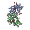 6a80S S: Starting model for refinement C: citing same article ( |
|---|---|
| Similar structure data |
-Links
 PDBj
PDBj- Assembly
| Deposited unit | 
| ||||||||
|---|---|---|---|---|---|---|---|---|---|
| 1 |
| ||||||||
| Unit cell |
|
-Components
| #1: Protein | Mass: 27615.590 Da / Num. of mol.: 2 Source method: isolated from a genetically manipulated source Source: (gene. exp.) Candidatus Liberibacter asiaticus str. psy62 (bacteria)Strain: psy62 / Gene: CLIBASIA_05070 / Production host: #2: Chemical | 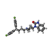  Mass: 461.546 Da / Num. of mol.: 2 / Source method: obtained synthetically / Formula: C28H29F2N3O / Feature type: SUBJECT OF INVESTIGATION Mass: 461.546 Da / Num. of mol.: 2 / Source method: obtained synthetically / Formula: C28H29F2N3O / Feature type: SUBJECT OF INVESTIGATION#3: Water | ChemComp-HOH / |  Mass: 18.015 Da / Num. of mol.: 246 / Source method: isolated from a natural source / Formula: H2O Mass: 18.015 Da / Num. of mol.: 246 / Source method: isolated from a natural source / Formula: H2OHas ligand of interest | Y | Has protein modification | Y | |
|---|
-Experimental details
-Experiment
| Experiment | Method: X-RAY DIFFRACTION / Number of used crystals: 1 |
|---|
- Sample preparation
| Crystal | Density Matthews: 4.31 Å3/Da / Density % sol: 71.49 % |
|---|---|
| Crystal grow | Temperature: 293 K / Method: vapor diffusion, sitting drop / Details: 0.1M Sodium citrate, 20% PEG 550, pH 5 |
-Data collection
| Diffraction | Mean temperature: 100 K / Serial crystal experiment: N |
|---|---|
| Diffraction source | Source: ROTATING ANODE / Type: RIGAKU MICROMAX-007 HF / Wavelength: 1.54 Å |
| Detector | Type: RIGAKU HyPix-6000HE / Detector: PIXEL / Date: Nov 17, 2021 |
| Radiation | Protocol: SINGLE WAVELENGTH / Monochromatic (M) / Laue (L): M / Scattering type: x-ray |
| Radiation wavelength | Wavelength: 1.54 Å / Relative weight: 1 |
| Reflection | Resolution: 2.65→28.8 Å / Num. obs: 29525 / % possible obs: 99.8 % / Redundancy: 4.3 % / CC1/2: 0.97 / Net I/σ(I): 5.5 |
| Reflection shell | Resolution: 2.65→9.01 Å / Num. unique obs: 3590 / CC1/2: 0.86 |
- Processing
| Software |
| |||||||||||||||||||||||||||||||||||||||||||||
|---|---|---|---|---|---|---|---|---|---|---|---|---|---|---|---|---|---|---|---|---|---|---|---|---|---|---|---|---|---|---|---|---|---|---|---|---|---|---|---|---|---|---|---|---|---|---|
| Refinement | Method to determine structure: MOLECULAR REPLACEMENT Starting model: 6A80 Resolution: 2.65→28.8 Å / Cor.coef. Fo:Fc: 0.879 / Cor.coef. Fo:Fc free: 0.821 / SU B: 12.172 / SU ML: 0.255 / Cross valid method: THROUGHOUT / σ(F): 0 / ESU R: 0.404 / ESU R Free: 0.299 / Stereochemistry target values: MAXIMUM LIKELIHOOD / Details: U VALUES : REFINED INDIVIDUALLY
| |||||||||||||||||||||||||||||||||||||||||||||
| Solvent computation | Ion probe radii: 0.8 Å / Shrinkage radii: 0.8 Å / VDW probe radii: 1.2 Å / Solvent model: MASK | |||||||||||||||||||||||||||||||||||||||||||||
| Displacement parameters | Biso max: 117.16 Å2 / Biso mean: 29.724 Å2 / Biso min: 2.49 Å2
| |||||||||||||||||||||||||||||||||||||||||||||
| Refinement step | Cycle: final / Resolution: 2.65→28.8 Å
| |||||||||||||||||||||||||||||||||||||||||||||
| Refine LS restraints |
| |||||||||||||||||||||||||||||||||||||||||||||
| LS refinement shell | Resolution: 2.65→2.719 Å / Rfactor Rfree error: 0 / Total num. of bins used: 20
|
